
Dr. Prangya Panda, P09876, Dr. Subudhi B N R, Dr. Sarita Panda, Dr. Suchitra dash
ABSTRACT
Human immunodeficiency virus is responsible for acquired immunodeficiency syndrome. It is a condition in humans in which progressive failure of the immune system allows life-threatening opportunistic infections and malignancies. Ocular lesions in the anterior segment of the eye may include blepharitis,molluscumcontagiosum, squamous cell carcinoma and Kaposi sarcoma,dry eye, herpes zoster ophthalmicus, uveitis, keratitis. Ocular lesions in the posterior segment of the eye include retinal vasculopathy, unusual malignancies, neuro ophthalmic lesions and opportunistic infections like cytomegalo virus (CMV) retinitis, toxoplasma choroiditis, syphilitic retinitis . The HIV positive cases examined were from medicine department, surgery department, obstetrics and gynecologydepartment, pulmonary medicine, antiretroviral therapy (ART)centre and patients attending ophthalmology OPD. With widespread introduction of ART in 1987, the ocular manifestations changed their pattern and prevalence. Most of the ocular complains were pain, redness, photophobia, lacrimation, defective vision, foreign body sensation andgrowth.
INTRODUCTION
Human immunodeficiency virus (HIV) is a lentivirus[1] (a member of the retrovirus[2] family) that causes acquired immunodeficiency syndrome (AIDS), a condition in humans in which progressive failure of the immune system[3] allows life-threatening opportunistic infections[4] and malignancies.
HIV infection in humans is considered by the World Health Organization (WHO), affecting about 65 million people worldwide (2008 study).India is the third largest population affected with AIDS accounting to 2-3 million population. HIV infects about 0.6% of the world’s population.
From its discovery in 1981 till 2006, death due to AIDS were more than 25 million. In 2004 itself, AIDS claimed 2.1 million lives which declined to 1.8 million in 2009. Treatment with antiretroviral drugs reduces both the mortality and morbidity of HIV infection. Although antiretroviral medication is still not universally available, expansion of antiretroviral therapy programs since 2004 has helped to turn the tide of AIDS deaths and new infections in many parts of the world. Intensified awareness and preventive measures, as well as the natural course of the disease, have also played a role in declining the morbidity.
Ocular lesions in the anterior segment of the eye may include blepharitis, dry eye, herpes zoster ophthalmicus, uveitis, keratitis, molluscumcontagiosum, squamous cell carcinoma and Kaposi sarcoma.[5]
Ocular lesions in the posterior segment of the eye include retinal vasculopathy, unusual malignancies, neuro ophthalmic lesions and opportunistic infections like CMV retinitis, toxoplasma choroiditis, syphilitic retinitis etc.[6]
With widespread introduction of ART in 1987, the ocular manifestations changed their pattern and prevalence. [7]
Our work aims to a comprehensive study of the incidence and various ocular manifestations in HIV positive patients.
METHOD
The study was conducted for a period of two years from November 2012 to October 2014 in M.K.C.G. Medical College and Hospital. All patients under study were known HIV positive cases. The present study comprises of all the HIV positive cases that had ocular manifestations. Out of which some were:
- Proved HIV cases attending Ophthalmology dept. with ocular problems.
Detailed ocular examination were done by Slit lamp biomicroscopy, Retinoscopy, Direct and Indirect ophthalmoscopy, Tonometry, Fluorescein staining, fundus photography and Perimetry. The medical records of the patients were used to obtain information on systemic diseases, HIV status, CD4 counts and use of antiretroviral therapy.All the cases were examined with proper precaution of using mask and gloves.
The name and registration number of the patient were kept with us, as record but was not revealed here, to keep the identity of the patient as a secret.
RESULT
INCIDENCE OF OCULAR MANIFESTATIONS IN HIV POSITIVE PATIENTS
TABLE 1
| Period of study | Total No. of cases | No. of cases with ocular manifestations | Percentage |
| Nov’12 – Oct’14 | 219 | 61 | 27.85% |
Though the number of patients on ART were more, but not all the patients turned to the Ophthalmology department for examination. Hence, 219 patients who attended the OPD were considered for the study .Out of which 61 cases were found to have ocular manifestations. As per the present study, the incidence of ocular manifestations in HIV patients was 27.85%
| Age in years | Total no. of cases with ocular manifestations | Percentage (n=61) |
| 0-10 | 1 | 1.64% |
| 11-20 | 2 | 3.28% |
| 21-30 | 13 | 22.95% |
| 31-40 | 29 | 47.54% |
| 41-50 | 13 | 19.67% |
| 51-60 | 2 | 3.28% |
| >60 | 1 | 1.64% |
| Total | 61 | 100% |
SEX DISTRIBUTIONAs per the present study, the incidence of ocular manifestations in HIV patients was found to be highest (47.54%) in the age group of 31-40 years. Mean age of presentation was 34.67 years. Second most common age group affected was between 21-30 years (22.95%).
TABLE-3
| Sex | No. of cases | Percentage(n=61) |
| Male | 45 | 72.13% |
| Female | 16 | 27.87% |
| Total | 61 | 100% |
Out of the 61 patients, 44 (72.13%) cases were male and 17 (27.87%) cases were female. In the study the male to female ratio was found to be 2.59:1
LATERALITY
TABLE-4
| Disease | Unilateral | Percentage
36.07% |
Bilateral | Percentage | |||
| Right eye | Left eye | Total
(n=61) |
Both eyes | Total
(n=61) |
|||
| HZO | 4 | 2 | 6 | – | 63.93% | ||
| Molluscum contagiosum | 1 | 1 | – | ||||
| Conjunctivitis | – | – | – | 5 | 5 | ||
| Blepharitis | – | – | – | 2 | 2 | ||
| Dry eye | – | – | – | 5 | 5 | ||
| Viral Keratitis | 1 | 1 | 2 | 1 | 1 | ||
| Ant. Uveitis | 1 | 1 | 2 | 1 | 1 | ||
| Kaposi Sarcoma | – | 1 | 1 | – | – | ||
| HIV retinopathy | 3 | 5 | 8 | 17 | 17 | ||
| CMV retinitis | – | – | – | 5 | 5 | ||
| Choroiditis | – | 2 | 2 | 1 | 1 | ||
| Papilledema
Optic atrophy |
– | – | – | 1
1 |
1
1 |
||
|
|
|||||||
| Optic atrophy | 1 | 1 | |||||
As per the present study, out of 61 cases, right eyes were involved in 10 (16.39%) cases and left eyes were involved in 12 (19.67%) amounting to a total of 22 (36.07%) cases of unilateral affection, whereas bilateral involvement was seen in 39 (63.93%) cases.
HZO always has a unilateral involvement whereas Conjunctivitis, Blepharitis,Dry Eye and retinopathies are mostly bilateral in presentation. Systemic association of Diabetes and Hypertension could also be a factor for bilateral presentation of retinopathies.
SOCIOECONOMIC STATUS
TABLE-5
| Socioeconomic status | No. of cases | Percentage (n=61) |
| Low | 40 | 65.57% |
| Middle | 9 | 14.75% |
| high | 12 | 19.68% |
| Total | 61 | 100% |
According to the present study, 40 (65.57%) cases belonged to low socioeconomic status (SES). Patients in high socioeconomic status (SES) group were next commonly affected population (19.68%) cases
The incidence of HIV is more among patients of either low or high socio economic status. It is comparatively less in middle socio economic status group. It could be due to lower education and awareness in patients belonging to lower socio economic status. They also stay away from home in some high risk areas for their occupation and earning.
The sophisticated lifestyle in higher socioeconomic status group may lead to increased incidence among this group. Some of the patients in this group also had Diabetes or Hypertension, which could be a contributory factor to the ocular manifestations.
LEVEL OF EDUCATION
TABLE-6
| Level of education | No. of cases | Percentage (n=61) |
| Less than class 7 | 35 | 57.38% |
| More than class 7 | 18 | 29.52% |
| Graduates and above | 8 | 13.11% |
| Total | 61 | 100% |
57.38% in the study had an education status below 7th standard. Most of the patients were from the rural areas of Southern Odisha were less educated. They belong to low socioeconomic status, so were more in favor of working to earn for their living rather than to seek education for themselves or for their children.
Wezi M Msishaet al (2003-04) in Tanzania observed that there was no marked association between increasing education and HIV sero prevalence.
The study is in agreement with the study done by Dr.NgonaBangaListo (2008).
MODE OF TRANSMISSION OF DISEASE
TABLE-7
| Mode of transmission | No. of cases | Percentage (n=61) |
| Sexual | 46 | 75.41% |
| Blood transfusion | 8 | 13.11% |
| Drug abuse | 2 | 3.28% |
| Needle prick injury | 3 | 4.92% |
| Mother to child | 2 | 3.28% |
| Total | 61 | 100% |
It is evident from the above table that the major route of HIV transmission (75.41%) is through sexual exposure, due to unprotected sex, uncommon methods of sex (oral sex). Parenteral route involving blood transfusion, needle prick injuries, accounts to the next most common method of transmission (21.31%) of HIV. Another 3.28% is due to transmission from mother to child.
PRESENTING COMPLAINS IN HIV POSITIVE PATIENTS UNDER STUDY
TABLE – 8
| Presenting symptoms | No. of cases (n=61) |
| Redness | 7 |
| Pain | 9 |
| Photophobia | 3 |
| Growth in either or both eyes | 2 |
| Defective vision | 18 |
| Foreign body sensation | 5 |
| Vesicles over the skin in peri-orbital lesion | 6 |
| Asymptomatic | 22 |
In my study, HIV positive patients were mostly referred from ART centre and other departments of M.K.C.G Medical College and Hospital who had no ocular problems. Next common group had defective vision, either for near or distance which is mostly due to refractive errors, retinopathies or lenticular opacities.
INCIDENCE OF OCULAR ADNEXAL MANIFESTATIONS IN HIV POSITIVE PATIENTS
TABLE – 9
| Adnexal manifestations | No. of cases | Percentage (n=61) |
| Herpes zoster Ophthalmicus | 6 | 9.83% |
| Blepharitis | 2 | 3.28% |
| Molluscum contagiosum | 1 | 1.64% |
| Total | 9 | 14.75% |
In the study, out of 61 HIV positive cases, 9 (14.75%) cases had adnexal manifestations and 6 cases presented with Herpes zoster Ophthalmicus, 2 with blepharitis and a single case of Molluscum contagiosum.
The HIV patients are immunocomprised, so are vulnerable to various other infections including herpetic infections. Patients with HZO had a prior history of Varicella affection and recurrent zoster infection Blepharitis got cleared with maintenance of proper lid and scalp hygiene, warm compresses, removal of lid debris with baby shampoo and antibiotic drops and ointments.
The lower incidence of Molluscum contagiosum could be due to less concern of the patients to take any medical help for such growths until they are symptomatic.
VISUAL ACUITY AT PRESENTATION IN HIV POSITIVE PATIENTS UNDER STUDY
TABLE-10
| VA | Right eye(n=61) | Left eye(n=61) |
| 6/6-6/12 | 47 | 46 |
| 6/18-6/36 | 9 | 11 |
| ³ 6/60 | 4 | 3 |
| Could not be recorded | 1 | 1 |
| Total | 61 | 61 |
Some of the patient’s visual acuity improved following treatment with topical drops and ointments (HZO, Keratitis), some improved following ART and T/t of systemic diseases.
Patients having diffuse exudates, intra retinal hemorrhages, associated systemic diseases (diabetic or hypertensive retinopathies), central choroiditis and optic atrophy presented with severe loss of vision.
INCIDENCE OF ANTERIOR SEGMENT MANIFESTATIONS IN HIV POSITIVE PATIENTS
TABLE-11
| Anterior segment manifestations | No. of cases | Percentage (n=61) |
| Bacterial Conjunctivitis | 5 | 8.20% |
| Dry eye | 5 | 8.20% |
| Viral Keratitis | 3 | 4.92% |
| Anterior Uveitis | 3 | 4.92% |
| Kaposi Sarcoma | 1 | 1.64% |
| Total | 17 | 27.88% |
From the above table, it is observed that, from the 17 (27.88%) cases with anterior segment finding out of 61 HIV positive patients, 5 (8.20%) cases were found to have dry eyes and the same number 5(8.20%) to have bacterial conjunctivitis. Rest included Keratitis 3 (4.92%), Uveitis 3 (4.92%) and Kaposi Sarcoma1(1.64%) cases. None were found to be having any Conjunctival microvasculopathyor Conjunctival Squamous cell carcinoma.
My study was comparable to the studies reported by other authors.
INCIDENCE OF POSTERIOR SEGMENT FINDING IN HIV POSITIVE PATIENTS
TABLE-12
| Posterior segment manifestations | No. of cases | Percentage (n=61) |
| HIV Retinopathy | 25 | 40.98% |
| CMV Retinitis | 5 | 8.20% |
| Choroiditis | 3 | 4.91% |
| Total | 33 | 54.09% |
As per my study, from among the 61 HIV positive patients, 33 (54.09%) cases have posterior segment findings and among them, the most common finding was HIV retinopathy in 25 (40.98%) cases. HIV retinopathy included, Cotton wool spots (CWS), intra retinal hemorrhages and retinal vascular micro aneurysms. Cotton wool spots were the most common finding in 22 (36.07%) cases. CMV Retinitis was found in 5 (8.20%) cases and rest 3 (4.91%) cases were having Choroiditis.
ART TREATMENT AND HIV POSITIVE PATIENTS
TABLE-14
| ART | No. of cases (n=61) | Percentage |
| Yes | 45 | 73.77% |
| No | 16 | 26.23% |
| Total | 61 | 100% |
Out of the 61 HIV positive patients, 45(73.77%) cases were on ART and rest 16 (26.23%) patients were not receiving any ART. The patients on ART were mostly referred from ART centre. The newly diagnosed HIV positive were those referred from other departments of our hospital for certain ocular ailments or simply for screening of any ocular manifestations.they were advised to attend the ART center for medical therapy.
SUMMARY AND CONCLUION
As per the study, the incidence of ocular manifestations was found to be 27.85% which was at par with other studies of various parts of the world.
The most common ocular manifestations were in the posterior segment of the eye 54.09%, followed by 27.88% in anterior segment. HIV retinopathy was the most common mode of presentation (40.98%).Cotton wool spots were found in 36.07% cases. This was followed by Herpes zoster Ophthalmicus 9.83% followed by conjunctivitis,Dry eye and CMV retinitis, each 8.20%, anterior uveitis, Viral Keratitis, and Choroiditis each of 4.92%; 3.28% cases of Blepharitis and 1.64% each of Molluscum contagiosum, Kaposissarcoma,Papilledema and Optic atrophy.[9] Hence, all the HIV positive patients should be screened for any ocular involvement as they may be asymptomatic but may have a posterior segment pathology which if not diagnosed at an early stage could lead to some blinding disease.
Periodically CD 4 Tcell should be monitored to record any decline in the CD 4 T cell count and immediate measures can be taken. [10]
HIV is a deadly disease taking tolls of life if untreated. Most of the HIV/AIDS patients present with ocular manifestations at some point of their disease process when the CD 4 Tcell count gradually declines below a critical level.
In the present study, most common ocular findings were HIV retinopathy of which Cotton wool spots were the most frequent finding.
Early diagnosis and starting of antiretroviral treatment can maintain CD4 T cell concentration, decrease the viral load and prevent various ocular complications and thus resulting morbidity contributed by HIV infection.
REFERENCE
- Lentivirus, https://en.wikipedia.org/wiki/Lentivirus
- Retrovirus, https://en.wikipedia.org/wiki/Retrovirus
- Immune system , https://en.wikipedia.org/wiki/Immune_system
- Opportunistic infection, https://en.wikipedia.org/wiki/Opportunistic_infection
- Sihotaramanjit, , Parsons diseases of the eye, 2016, 22nd edition, 537 page
- Principles and practices of ophthalmology by peyman , sanders , and Goldberg
- Yanoff and duker ophthalmology
- Samarbasak,Essentials of ophthalmology, 2nd edition, 1999, 90 page
- Khurana A K, comprehensive ophthalmology, 6th edition, 2015,469 page
- Jogi Renu, basic ophthalmology, 3rd edition, 2003, 389 page
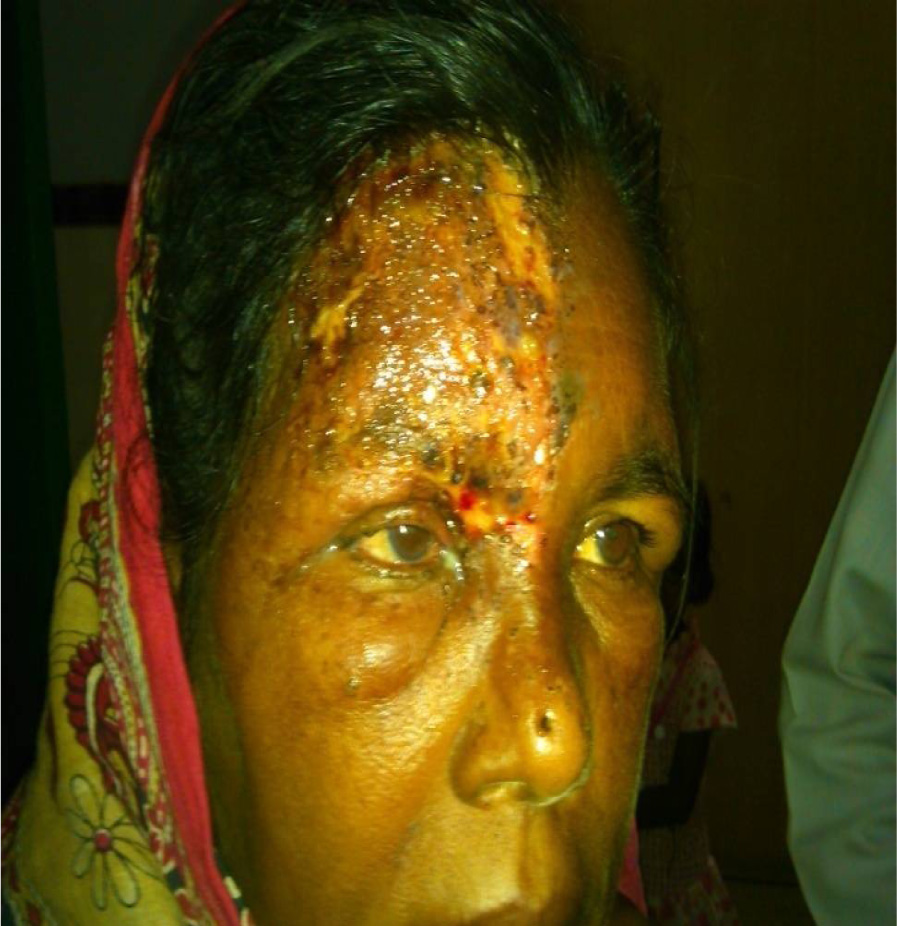
Herpes zoster ophthalmicus in a case of hiv
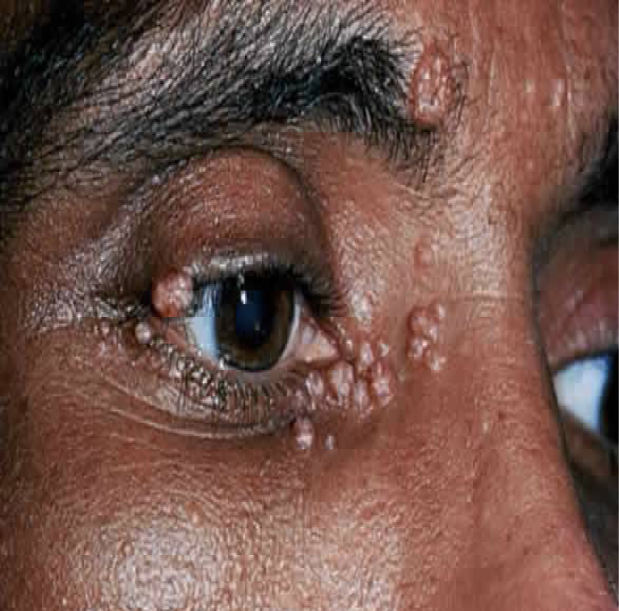
Molluscum contagiosum in a case of hiv
Leave a Comment