
Dr. Anushri Agrawal, A18605, Dr. Rengaraj Venkatesh, Dr. Kavitha S, Dr. Swati Upadhyaya
Introduction
Glaucoma is a chronic, progressive optic neuropathy caused by a group of ocular conditions, which lead to damage of the optic nerve with loss of visual function.[1]
According to the World Health Organization, glaucoma is now the leading cause of blindness in the world, second only to cataracts, and is considered the principal cause of irreversible blindness in the world[2]with a higher morbidity among women and Asians.[3-5]
Unfortunately, it is estimated that only half of glaucoma sufferers are diagnosed and treated at present time[6-8] and the number of undiagnosed glaucoma patients could be much higher in developing or undeveloped countries, reaching more than 90% in rural India.[9]
Using a non-mydriatic fundus camera is a reasonable way to improve access to high-quality glaucoma screening. However, most previously tested cameras are tabletop models limiting their portability and applicability in rural outreach settings, especially in the developing world. The objective of this study is to evaluate the sensitivity and specificity of a handheld non-mydriatic fundus camera to aid in the diagnosis of glaucoma. However, the first step towards evaluating the possible use of a diagnostic tool as a screening method is defining its accuracy for the diagnosis of the disease. The purpose of our study is to evaluate the accuracy of a set of criteria in evaluating the optic nerve head in non-mydriatic fundus photography for the diagnosis of glaucoma.
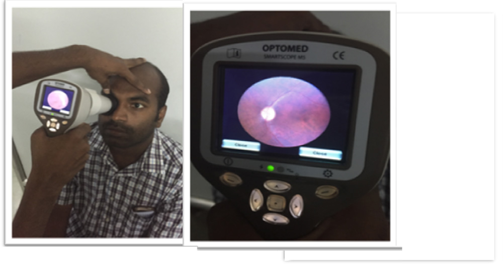
Optomed Smartoscope M5
Optomed Smartscope M5 is a digital medical camera that provides general, ophthalmoscopic, otoscopic and dermatoscopic imaging with one hand-held device. This multipurpose digital imaging device weights only 400g and powered with a battery it gives you the freedom to move around and take the device with you to any location.
Digital still and video images created with Optomed Smartscope M5 allow making accurate first diagnosis and planning consistent follow-up treatment.
 Materials and Methods:
Study Area:
Glaucoma services of Aravind Eye Hospital, Pondicherry.
Study Design:
Cross-sectional study.
Study Population:
Patients diagnosed with primary glaucoma that satisfy the inclusion and exclusion criteria were included in the study as cases and patients visiting general outpatient department who satisfy inclusion criteria for controls served as the control arm.
Sample Size:
138 eyes with primary glaucoma and 138 eyes of normal subjects.
Inclusion Criteria for Cases
- Age >30 years and <70 years.
- Diagnosed with POAG/PACG/ NTG with the relevant field defect who are under medical or post surgical management.
- Willing to give written informed consent.
Inclusion Criteria for Controls
- Age >30 years and <70 years.
- Not diagnosed/suspect for glaucoma.
- Negative family history.
- Cup disc ratio of <=0.5 and inter eye asymmetry of less than 0.2.
- Myopia or hypermetropia of less than 3D, presbyopia.
- Willing to give written informed consent.
Exclusion Criteria
- Secondary glaucoma such as PEXG or PG; angle recession and traumatic glaucomas, intraocular neovascularization, and neovascular glaucoma.
- Cataract or media opacity including Posterior capsular opacification (PCO) which interferes with fundus imaging, cataracts of grade more than NC3, NO3, C3 or P3 as per LOCS III classification.
- Vitreo-retinopathy of any etiology except mild to moderate Non Proliferative Diabetic Retinopathy.
- Neurological damage to disc – traumatic optic neuropathy / optic neuritis.
Informed consent was obtained from patients to participate in study and choice to decline participation was made available. The protocol was approved by the Institutional Review Board of Aravind Eye Hospital and Post-Graduate Institute of Ophthalmology.
Patients presenting to the Glaucoma service / Outpatient department of Aravind eye hospital, Pondicherry
Preliminary examination by Glaucoma specialist
Undilated images of optic disc, 45° from each eye using the smartscope (Optomed, Finland) by a single technician
Pupils dilated
Gold standard disc evaluation by senior Glaucoma consultant
Images digitalized and de-identified by an independent investigator
Images of patients and controls presented to the two masked graders randomly for grading glaucomatous changes in sets of 200 images
Re-identification of the images and tabulation of results
Sensitivity and specificity of undilated Optomed images will be calculated by comparing with the gold standard clinical examination with 90 D lens.
The observers were asked to evaluate each image for the presence of the following criteria of glaucomatous optic nerve damage:
- Vertical C/D ratio (Figure 1)
- Notching or thinning of the neuroretinal rim (Figure 1)
- Retinal nerve fibre layer defect (Figure 2)
- Disc hemorrhages (Figure 3)
- Peri papillary atrophy (Figure 4)
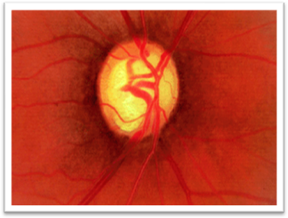
Figure 1: Glaucomatous cupping with inferior NRR notching
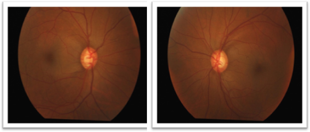
Figure 2: Retinal nerve fibre layer defects depicted in our patients
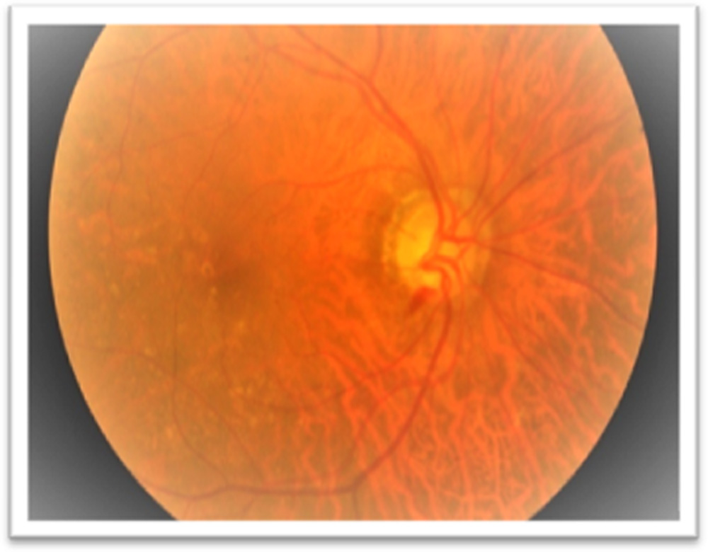
Figure 3: Splinter hemorrhage taken by Optomed camera
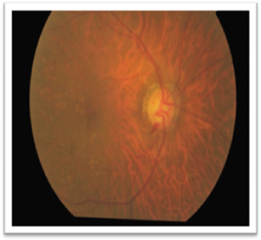
Figure 4: Peri papillary atrophy on fundus photograph
Appropriate statistical test like mean, SD, range were applied for descriptive purpose and for drawing inference of collected data- tests of significance like paired and unpaired t test, Chi square test, Bland Altmann analysis were applied with the help of biostatistician.
Results
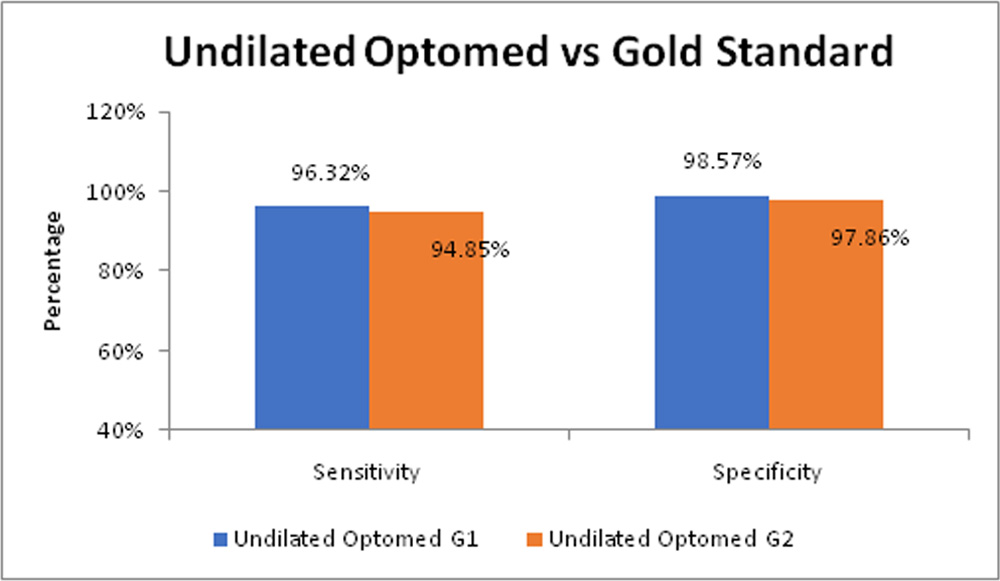
- The sensitivity and specificity of undilated Optomed fundus camera as compared to +90D clinical examination was found to be 96.32% (91.6 – 98.8%) and 98.57% (94.9 – 99.8%) respectively for grader 1 (G1) and 94.85% (89.7 – 97.9%) and 97.86% (93.9 – 99.6%) respectively for grader 2 (G2).
Table 1: Undilated Optomed G1 vs Gold Standard
| Undilated Optomed-G1 | Gold Standard | Sensitivity & Specificity (CI) | |
| Yes | No | ||
| Yes | 131 | 2 | Sensitivity: 96.32% (91.6 – 98.8%)
Specificity: 98.57% (94.9 – 99.8%) |
| No | 5 | 138 | |
| Total | 136 | 140 | |
Table 2: Undilated Optomed G2 vs Gold Standard
| Undilated Optomed-G2 | Gold Standard | Sensitivity & Specificity (CI) | |
| Yes | No | ||
| Yes | 129 | 3 | Sensitivity: 94.85% (89.7 – 97.9%)
Specificity: 97.86% (93.9 – 99.6%) |
| No | 7 | 137 | |
| Total | 136 | 140 | |
Figure 5: Undilated Optomed vs Gold Standard
Conclusions
- According to our study results, the non mydriatic fundus camera has a high sensitivity and specificity (of more than 90%) in detecting glaucoma as compared to gold standard +90D examination.
- The fundus photographs taken by hand-held Optomed fundus camera are comparably effective (greater than 90% sensitivity and specificity) in screening glaucoma as compared to photographs taken by the standard table-top Topcon fundus camera.
- Hence non mydriatic fundus photography can be an valuable tool for screening glaucoma especially useful in outreach programmes.
References
- Sihota R, Tandon R. The Glaucoma. In: Sihota R, Tandon R, editors. Parsons’ Disease of the eye. 21st ed. India: Elsevier; 2011. p. 280–300.
- Resnikoff S, Pascolini D, Etya’ale D, Kocur I, Pararajasegaram R, Pokharel GP, et al. Global data on visual impairment in the year 2002. Bull World Health Organ. 2004;82(11):844–51.
- Quigley HA. Number of people with glaucoma worldwide. Br J Ophthalmol. 1996;80(5):389–93.
- Thylefors B, Negrel AD. The global impact of glaucoma. Vol. 72, Bulletin of the World Health Organization. 1994. p. 323–6.
- Quigley HA, Broman AT. The number of people with glaucoma worldwide in 2010 and 2020. Br J Ophthalmol. 2006;90(3):262–7.
- Yip JLY, Foster PJ. Ethnic differences in primary angle-closure glaucoma. Curr Opin Ophthalmol. 2006;17(2):175–80.
- Tielsch JM, Sommer A, Katz J, Royall RM, Quigley HA, Javitt J. Racial variations in the prevalence of primary open-angle glaucoma. The Baltimore Eye Survey. JAMA J Am Med Assoc. 1991;266(3):369–74.
- Weih L. Prevalence and predictors of open-angle glaucoma Results from the visual impairment project. Ophthalmology. 2001 Nov;108(11):1966–72.
- Lee BW, Sathyan P, John RK, Singh K, Robin AL. Predictors of and Barriers Associated With Poor Follow-up in Patients With Glaucoma in South India. Arch Ophthalmol. 2008 Oct 13;126(10):1448.
Leave a Comment