
Dr. Madhusudan MandaL, M19430, Dr. Rizvi Syed Ali Raza, Dr. R Maheshwari
Abstract
Objective: To evaluate the level of Interleukin-4 (IL-4)in tears of seasonal allergic conjunctivitis (SAC) and vernal keratoconjunctivitis (VKC) patients with a hope to discover new insights into the immunopathogenesis of allergic conjunctivitis that would lead to the development of more effective pharmacologic agents in the treatment of allergic conjunctivitis.
Material and methods: Tears samples were collected from 16 patients of SAC, 16 patients of VKC and 10 healthy controls. IL-4 levelswere measured in tears using Enzyme-linked Immunosorbent Assay (ELISA) test.Patients having any non-ocular allergy, dry eye and other ocular inflammatory disease or any autoimmune inflammatory disease are excluded from the study.
Results: IL-4 measurement in tears would be a fair test to discriminate SAC and VKC patients from controls and cut off value of IL-4 would be 1.07 pg/ml in SAC and 0.93 pg/ml in VKC. The mean level of IL-4 in tears of VKC patients was significantly higher than that of controls (p = 0.011) and that of SAC patients (p = 0.022).
Conclusions: IL-4 is an important pro-inflammatory marker in the immunopathogenesis of SAC and VKC. IL-4 could be a diagnostic marker as well as a therapeutic target in these diseases.
Keywords:seasonal allergic conjunctivitis, vernal keratoconjunctivitis,interleukin-4,
enzyme-linked immunosorbent assay
Introduction
Allergic conjunctivitis (AC) is a disease primarily characterized by an inflammatory response of the conjunctival mucosa.[1]It is a hypersensitivity response to external antigens, usually called allergens.[2] AC is not a single disease; in fact it is a syndrome affecting the entire ocular surface, including conjunctiva, lids, cornea, and tear film.[3]
Allergic conjunctivitis is one of the most common ocular conditions encountered in clinical practice. It includes a spectrum of different clinical entities with variable presentation. AC is classified into mild forms, such as seasonal allergic conjunctivitis (SAC), perennial allergic conjunctivitis (PAC) as well as giant papillary conjunctivitis (GPC), and more severe, chronic forms such as vernal keratoconjunctivitis (VKC) and atopic keratoconjunctivitis (AKC).[4]The different types of AC provide a fascinating insight into the various immune mechanisms affecting the conjunctiva.
As the prevalence of allergic disease is increasing, probably through environmental factors, it is appropriate at this stage to study the immunopathogenesis of allergic conjunctivitis.SAC is caused by immunoglobulin E (IgE) mediated mast cell degranulation whereas in VKC there is continuous over-expression of mast cells, eosinophils, neutrophils, fibroblast and lymphocytes which is mediated by allergen specific IgE response and predominantly by T helper type 2 (Th2) cells in a complex pathogenesis.[5] The degranulated mast cells release inflammatory mediators like histamine, prostaglandins, leukotrienes, interleukins which finally produce inflammations.
With the growing body of literature, the involvement of cytokines in the recruitment and activation of inflammatory cells in AC is being more apparent. IL-4 regulates biological responses by binding to specific IL-4 receptors (IL-4Rs) expressed by mast cells, eosinophils, basophils and abundant activated lymphocytes[6] which are found in conjunctival histopathological examination of AC patients. In this study, we had evaluated the level of IL-4 in tears of SAC and VKC patients using Enzyme-linked Immunosorbent Assay (ELISA) system and compared those with control group with a hope to discover new insights into the immunopathogenesis of the allergic conjunctivitis that would lead to the development of more effective pharmacologic agents in the treatment of allergic conjunctivitis.
Material and Methods
This prospective cohort study was conducted over 1-year duration in the department of Ophthalmology at a tertiary-level teaching hospitalin accordance with the ethical standards formulated in the Helsinki Declaration. Informed written consent was taken from all the patients or their guardians (in case of minor).32 patients were enrolled in this study, of which 16 patients were in SAC group and 16 in VKC group.The patients of SAC and VKC were diagnosed on the basis of clinical signs and symptoms. 10 age and sex matched healthy individuals served as control group.Patients having any non-ocular allergy, dry eye and other ocular inflammatory disease or any autoimmune inflammatory disease were excluded from the study.
Sample collection and storage
A volume of tears exceeding 100 µl was collected from single eyes of the subjects with a micropipette (Thermo Fischer Scientific) and disposable microtips (Tarsons) and transferred into Eppendrof tubes -1.5ml (Future Bio Science). Tears samples were stored at -20ºC until assayed.
Measurement of IL-4
IL-4 was measured by the ELISA method (Diaclone Human IL-4 ELISA kit, France) according to the manufacturer’s instructions. The absorbance value (OD value) of each well was read on a spectrophotometer at a wavelength of 450 nm. We then drew the standard curve which was almost linear and calculated the concentrations of IL-4 by extrapolating OD values against IL-4 standard concentrations using the standard curve.
Statistical analysis:
We analysed the data in our study using SPSS statistical software (version 20).Demographic characteristics were presented as mean ± standard deviation (SD) for continuous variables and as frequencies and percentages for categorical variables.IL-4 values were reported as mean ± SD (pg/ml). The mean IL-4 levels were compared between the different groups using unpaired two-tailed Student’s t-test. The association between IL-4 level and disease duration was detected using Pearson correlation analysis. All analyses in this study employed a significance level of p < 0.05 (two-tailed).
A receiver-operating characteristic (ROC) curve was used to describe the performance of continuous variable. The predictive accuracy of IL-4 levels in patients with SAC and VKC was examined using the area under the ROC (AUC). Optimal cut-off values were determined from ROC curve analyses by calculating the minimum distances from graph coordinates with maximum sensitivity and specificity.
Results
The mean age of the patients in SAC group was 14.2 ±3.68 years (range 9 to 21). In VKC and Control group, it was 11.5 ±3.09 years (range 8 to 17) and 11.6 ±2.87 years (range 8 to16) respectively. So the patients of SAC were older than the patients of VKC which was statistically significant (p = 0.031). Twelvepatients (75.0%) in SAC group and 10 patients (62.5%) in VKC group were in the age distribution of 10 to <20 years. Only 2 patients of SAC were in the age group ≥20 years. Thus the incidence of allergic conjunctivitis was highestin the age group of 10 to <20 years (68.75%).In this study, 11 patients (68.75%) in SAC group and12 patients (75%) in VKC group were male. Hence males were more affectedthan female.
The mean level of IL-4 in tears of control group was 1.02 ± 0.40 pg/ml (median 1.04 pg/ml and range being 0.94 pg/ml to 1.06 pg/ml) while that in SAC and VKC groups were 1.62 ± 1.33 pg/ml (median 1.20 pg/ml and range being 0.87 pg/ml to 6.16 pg/ml) and 3.53 ± 2.87 pg/ml (median 1.86 pg/ml and range being 0.93 pg/ml to 10.38 pg/ml) respectively (Figure 1).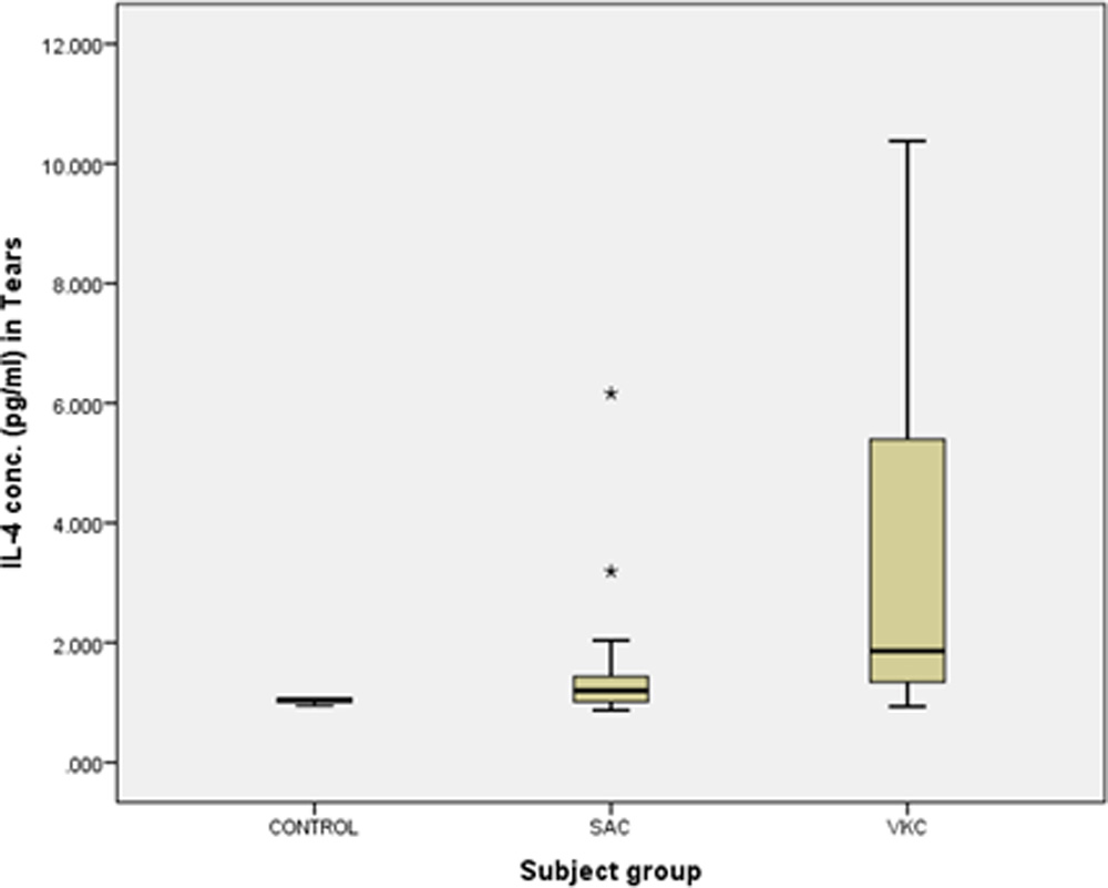
Figure1. IL-4 concentration in tears of subject groups.
However there was no significant difference in IL-4 level in tears between patients of SAC and controls (p = 0.170) but the ROC curve analysis revealed that IL-4 measurement in tears would be a fair test to discriminate SAC patients from controls (AUC = 0.713) and cut off value of IL-4 in tears to diagnose SAC would be 1.07 pg/ml (sensitivity: 62.5% and specificity: 100%) (Figure2).
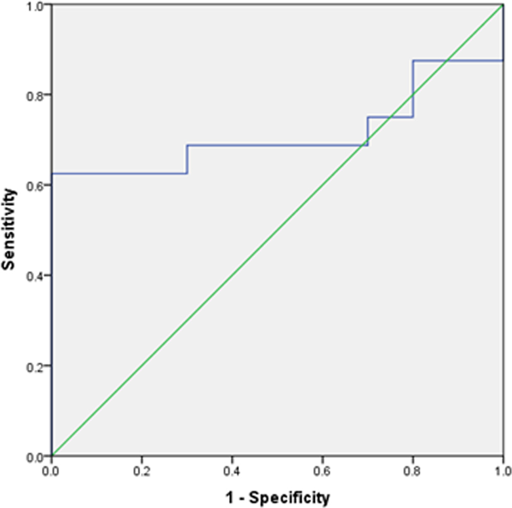
Figure2. ROC curve for IL-4 concentration in tears in SAC patients as compared to controls
The mean level of IL-4 in tears was significantly higher in patients of VKC as compared to control group (p = 0.011). ROC curve also revealed that IL-4 measurement in tears would be an excellent test to discriminate VKC patients from controls (AUC = 0.938) and cut off value of IL-4 in tears to diagnose VKC would be 1.162pg/ml (sensitivity: 93.8% and specificity: 100%) (Figure3).The mean IL-4 level in tears was significantly higher in VKC patients than SAC patients (p = 0.022). Another important finding found in this study was significant correlation between IL-4 levels and duration of disease (SAC, r =564 and p = 0.023; VKC, r = 0.583 and p = 0.018).
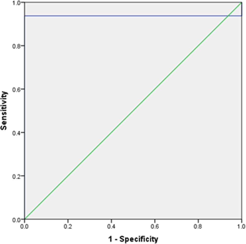
Figure3. ROC curve for IL-4 concentration in tears of VKC patients as compared to controls.
Discussion
Isolation and validation of the specific biomarkers reflecting the pathological changes in AC have major importance in therapeutics. The pleiotropic impact of interleukin-4 (IL-4) on immune system cells and its association with T helper type 2 (Th2) cells suggest that IL-4 might be involved in the pathogenesis of AC.[7]The previous studies showed the expression of the interleukin-4 receptor α in human conjunctival epithelial cells. Matsuura N et al.showed that the frequency of IL-4-producing conjunctival CD4+ T cells in patients with VKC were significantly higher than control.[8] Fujishima H et al. observed the significantly elevated levels of IL-4 in the conjunctival cells supernatants in allergic conjunctivitis.[9]
In our study, the mean IL-4 level in tears of control group was 1.02±0.40 pg/ml while that in SAC and VKC groups were 1.62 ±1.33 pg/ml and 3.53 ± 2.87 pg/ml respectively. H Fujishima et al.did a study where the mean level of IL-4 in tears of the normal control was 0.92± 0.59 pg/ml whereas that in the patients of SAC and VKC were 3.51 ± 2.53 pg/ ml and 23.95± 22.52 pg/ml respectively.[10] The less number of patients with severe form, different environmental factors and different subjects profile including socioeconomic status might be the causes of low IL-4 value in tears of allergic patients in our study.
Though the Post Hock (Tukey HSD) analysis showed that there was no statistically significant difference in IL-4 concentration in tears between patients with SAC and control (p = 0.170), the ROC curve analysis revealed that IL-4 measurement in tears would be a fair test to discriminate SAC patients from controls (AUC= 0.713) and the cut off value of IL-4 in tears to diagnose SAC would be 1.07 pg/ml (sensitivity: 62.5% and specificity: 100%). The mean IL-4 level in tears of VKC patients was significantly higher than that of controls (p = 0.011). The ROC curve also revealed that IL-4 measurement in tears would be an excellent test to discriminate VKC patients from controls (AUC = 0.938) and cut off value of IL-4 in tears to diagnose VKC would be 1.162pg/ml (sensitivity: 93.8% and specificity: 100%). So IL-4 level in tears of the patients might be a diagnostic marker in both VKC and SAC. Leonardi A et al. also found the elevated level of IL-4 in human tear specimens in seasonal and chronic allergic eye disease.[11] Fujishima et al.conducted a study where they found that the levels of IL-4 (p = 0.01) and histamine (p = 0.02) were significantly increased in the specimens of patients with cedar pollen-allergic conjunctivitis than postsurgical conjunctivitis.[12]
The mean IL-4 level in tears was significantly elevated in VKC groups as compared to SAC group (p=0.022). So VKC would be more severe form of allergic eye disease than SAC and IL-4 level measurement in tears of the patients might be indicated to differentiate VKC from SAC.
The positive correlation between IL-4 levels and duration of disease pointed out that both SAC and VKC would be considered as progressive disease. Hence the patients should be advised to take early treatment to prevent the progression.
Conclusions
These results indicate that IL-4 would bean important pro-inflammatory marker in the pathogenesis of SAC and VKC. So IL-4 would be a therapeutic target and IL-4 level in tears might be a diagnostic marker in these diseases.
Suggestions
The study was limited due to its sample size. Another replicative study in a larger cohort is required.
Financial support and sponsorship
Nil.
Conflicts of interest
There are no conflicts of interest.
References
- Origlieri C, Bielory L. Emerging drugs for conjunctivitis. Expert Opin Emerg Drugs. 2009;14:523–36.
- Friedlaender MH. Current concepts in ocular allergy. Ann Allergy 1991; 67:5-13.
- Johansson, SG., Bieber, T., Dahl, R., Friedmann, PS., Lanier, BQ., et al. (2004). Revised nomenclature for allergy for global use: Report of the Nomenclature Review Committee of the World Allergy Organization, October, 2003. J Allergy Clin Immunol. pp. 832-6.
- Messmer EM. Ocular allergies. Ophthalmologe. 2005 May;102(5):527-43; quiz 544.
- Leonardi A, De Dominicis C, Motterle L: Immunopathogenesis of ocularallergy: schematicapproach to different clinical entities. Curr Opin Allergy Clin Immunol 2007
- Vita, N, Lefort S, Laurent P, Caput D, Ferrara P. Characterization and comparison of theinterleukin 13 receptor with the interleukin 4 receptor on several cell types. J. Biol. Chem., 1995. 270: 3512–3517.
- Leonardi A1, Fregona IA, Plebani M, Secchi AG, Calder VL. Th1- and Th2-type cytokines in chronic ocular allergy. Graefes Arch Clin Exp Ophthalmol. 2006 Oct;244(10):1240-5.
- Matsuura N, Uchio E, Nakazawa M, Yago T, Matsumoto S, Ohno S, Minami M. Predominance of infiltrating IL-4-producing T cells in conjunctiva of patients with allergic conjunctival disease. Curr Eye Res. 2004 Oct-Nov;29(4-5):235-43.
- Fujishima H, Saito I, Takeuchi T, Shinozaki N, Tsubota K. Measurement of interleukin-4 and histamine in superficial cells of conjunctiva in patients with allergic conjunctivitis. Curr Eye Res. 1996 Feb;15(2):209-13.
- H Fujishima, T Takeuchi, N Shinozaki, I Saito, and K Tsubota.Measurement of IL-4 in tears of patients with seasonal allergic conjunctivitis and vernal keratoconjunctivitis. Clin Exp Immunol. 1995 November; 102(2): 395–398.
- Leonardi A, Curnow SJ, Zhan H, Calder VL. Multiple cytokines in human tear specimens in seasonal and chronic allergic eye disease and in conjunctival fibroblast cultures. Clin Exp Allergy. 2006 Jun;36(6):777-84.
- Fujishima H, Saito I, Takeuchi T, Shinozaki N, Tsubota K. Measurement of interleukin-4 and histamine in superficial cells of conjunctiva in patients with allergic conjunctivitis. Curr Eye Res. 1996 Feb;15(2):209-13.
Leave a Comment