
Dr. Sachin Arya, A15092, Dr. Bhushan Ghodke, Dr. Ashok Kumar Meena, Dr.Parmar Gautam Singh
ABSTRACT:
AIM: To evaluate outcomes of autologous simple limbal epithelial transplant (SLET) with amniotic membrane transplant(AMT) in children with unilateral chemical injury.
METHODS: Retrospective,case series of all children <18 years of age who have undergone autologous SLET with AMT procedure in unilateral chemical injury.
RESULTS: 24 eyes of 24 children (Male:Female -3:1) with mean age of 11.9 ± 5.7 years. All patients were between Grade 5 and 6 according to Dua’s Classification (Kindly mention classification in brief). Mean Follow-up was 8 ± 4.4 months. Mean time to complete epithelialization was 28.4 ± 8.9 days.Complications were encountered in 2 patients. Anatomical success was seen in 81.72% patients and visual success was noted in 43.54% patients.
CONCLUSIONS:SLET with AMT is a feasible option in children with severe unilateral chemical injury with fair anatomical and visual outcome.
INTRODUCTION:
Chemical injuries to the eye represent an ophthalmic emergency that can result in extensive damage and significant ocular morbidity. The reported incidence of ocular chemical injuries in developing countries is approximately 1.25% to 4.4%. Severe chemical burn can lead to complete destruction of the ocular surface, corneal opacification, permanent vision loss, and rarely loss of the eye.Alkalis cause significantly greater damage compared with acids. Majority of these injuries occur as a result of accidents at workplace or home or deliberately from an assault.
Early management involves preservation of globe integrity while in chronic phase, management of limbal stem cell deficiency come into play. Recently, a novel surgical technique was described, which claimed to combine the advantages of both CLAU (MENTION FULLFORM OF CLAU) and CLET, while eliminating the major drawbacks of both
techniques. In simple limbal epithelial transplantation (SLET), a small piece of donor limbal tissue is harvested and cut into multiple pieces and placed on the recipient surface, using human amniotic membrane (hAM) to support in-vivo expansion of epithelial cells.
Thus, the purpose of our study was to evaluate the outcome of SLET with AMT in pediatric chemical injury in a tertiary care centre.
METHODS:
It was a retrospective clinical non-comparative study conducted at SadguruNetraChikitsalaya, Chitrakoot. Medical records of all patients less than 18 years of age with history of ocular chemical injury and had undergone treatment with SLET in our centre were reviewed. The study was approved by an institutional review board and adhered to the tenets of the Declaration of Helsinki. All children were graded according to Dua’s classification for ocular chemical injury. Inclusion criteria included consecutive cases of unilateral LSCD with wet ocular surface for which autologous SLET was performed, with a minimum of 6 months of postoperative follow-up. Cases of LSCD secondary to immune mediated conditions such as Stevens–Johnson syndrome, mucous membrane pemphigoid and those with dry ocular surfaces were excluded. The diagnosis of LSCD was based on clinical signs such as absence of pigmented palisades of Vogt, irregular and lustreless corneal epithelium, persistent epithelial defects, fibrovascularpannus formation and conjunctivalisation of the corneal surface. Demographic details, aetiology of LSCD, prior surgery performed and clinical details including visual acuity at presentation, extent of LSCD (in clock hours), presence or absence of eyelid abnormalities, symblepharon and persistent epithelial defects were noted.
The surgical technique of SLET has been described previously by its respective authors.In brief, a small piece of limbal tissue (1–2 clock hours) was harvested from the unaffected eye. Fibrovascular pannus was excised from the eye with LSCD, and hAM was spread over the bare surface, using fibrin glue as adhesive. Significant symblepharon, if present, was released and the bare area covered by placement of a conjunctival autograft. The limbal tissue was cut into multiple small pieces (typically 10–15), which were distributed over the hAM with application of more fibrin glue and covered with a bandage contact lens (BCL) or another piece of hAM. Postoperatively, topical antibiotic (5% Moxifloxacin) eye drops were prescribed till removal of the contact lens or complete epithelisation of the surface. Topical steroid (1% Prednisolone acetate) eye drops were prescribed in tapering doses over 4–6 weeks. The primary outcome measure was clinical success, defined as a completely epithelised, avascular, stable corneal surface. Failure was defined as a recurrence of fibrovascular pannus encroaching on the central cornea, frequent epithelial breakdown or persistent epithelial defects. Continuous parametric data were reported as mean (± standard deviation) and nonparametric data were reported as median with range.Success rates were reported as percentages with 95% confidence intervals (CI).Cox proportional hazards analysis assessed association of preoperative characteristics with risk of failure.P value less than 0.05 was considered statistically significant.
RESULTS:
A total of 24 eyes of 24 children (Male:Female – 3:1) had undergone SLET procedure for limbal stem cell deficiency secondary to ocular chemical injury.
The mean age of patients was 11.9 ± 5.7 years (4 year – 16 years). All patients suffered ocular chemical injury due to bursting of chuna packets at home or outside with accidental entry of lime particles into eyes.The median duration from original injury to SLET was 22 Months (15 days – 7 years). Out of 24 children, 20 patients belonged to grade 5 Dua’s classification while 4 patients were graded as grade 6 of Dua’s classification. A completely epithelised, avascular corneal surface (clinical success) was achieved in 83.3% of cases. The mean period of complete epithelialization was 28.4 ± 8.9 days (range ??). 2 patients did not achieve avascular surface and eventually failed within 3 months following SLET.In the Cox proportional hazards analysis, presence of symblepharon (HR 9.4), loss of BCL (HR 20.8) and presence of infection (HR 7.8) were found to be significantly associated with a risk of failure of SLET. Pre-operatively 18 eyes had a visual acuity of perception of light and accurate projection of rays in all quadrants, of which 12 eyes improved to 3/60 at 6 months post-SLET (18 eyes gained vision, 2 eyes failed; what about the visual recovery of remaining 4 patients?). Recurrence of focal pannus was the most common complication followed by infection.
DISCUSSION:
The overall success rate of SLET in this study was 83.3% which remained above 80% at the final follow-up of 6 months. This is similar to or better than most published results of CLAU or CLET. In their initial report of direct limbalautograft transplantation, Keivyon and Tseng (reference number?) reported ‘stable epithelial adhesion’ in 20 of 21 cases, arrest or regression of corneal neovascularisation in 15 cases and improvement in visual acuity in 17 cases. Using combination of autolimbal and allolimbal sources, Shimazaki et aland Miri et al(reference number?) reported long-term success rates of 53.1% and 82%, respectively, with direct limbal transplantation. Despite the encouraging success rates with CLAU, the amount of donor limbal tissue required (10–20 mm or up to 6 clock hours) remains a concern. Using in vivo confocal microscopy to assess donor eyes used for harvesting limbal transplants, Miriet alfound the re-epithelised donor site to be covered by conjunctival epithelium in a large number of cases. By harvesting such a large amount of limbal tissue, there exists a potential for inducing iatrogenic LSCD at the donor site in CLAU. In contrast to this, SLET uses very little donor limbal tissue, minimising the area at risk for LSCD in the donor eye. More recently, a meta-analysis of 572 eyes found a success rate of 67% for CLET, with no difference between results of autologous and allogeneic transplants (REFERENCE?).There are a few fundamental differences between SLET and CLET. As the need for ex-vivo expansion of cells is obviated, SLET can be offered virtually anywhere by a trained surgeon at a fraction of the cost of CLET, the availability of which is restricted to very few centres. Our results suggest that spreading donor limbal tissue
over the cornea results in outcomes that are equally good or better than those achieved by placing it near the recipient limbus. Rates of improvement in visual acuity in this study are comparable with those reported for CLET. Baylis et aland Zhao et aldescribed an improvement of two or more lines of visual acuity in 51% and 67% of eyes, respectively, with CLET. One of the common complication encountered in this study was focal recurrence of LSCD. Such recurrences may be safely observed in case they are non-progressive and do not threaten the visual axis. In case intervention is required, a repeat SLET using a minimal amount of limbal tissue can be safely performed to specifically address the areas where pannus has recurred.
In conclusion, multicentre results indicate that autologous SLET is an effective and safe modality for treatment of unilateral LSCD. Clinical success rates and gain in visual acuity are equal to or better than those reported with CLET. In addition, it is a single-stage procedure, does not require expensive laboratory facilities for ex-vivo cell cultivation and can be easily learnt by cornea surgeons.
REFERENCES:
- Tseng SC. Concept and application of limbal stem cells. Eye (Lond) 1989;3(Pt 2): 141 –57.
- Dua HS, Saini JS, Azuara-Blanco A, et al. Limbal stem cell deficiency: concept,aetiology, clinical presentation, diagnosis and management. Indian J Ophthalmol2000;48:83–92.
- Keivyon KR, Tseng SCG. Limbalautografttransplantation for ocular surface disorders. Ophthalmology 1989;96:709–23.
- Daya SM, Chan CC, Holland EJ, et al. Cornea Society nomenclature for ocular surface rehabilitative procedures. Cornea 2011;30:1115–19.
- Pellegrini G, Traverso CE, Franzi AT, et al. Long-term restoration of damaged corneal surfaces with autologous cultivated corneal epithelium. Lancet 1997;349:990–3.
- Sangwan VS, Basu S, MacNeil S, et al. Simple limbal epithelial transplantation (SLET): a novel surgical technique for the treatment of unilateral limbal stem cell deficiency. Br J Ophthalmol 2012;96:931 –4.
- Vazirani J, Basu S, Sangwan V. Successful simple limbal epithelial transplantation (SLET) in lime injury-induced limbal stem cell deficiency with ocular surfacegranuloma. BMJ Case Rep 2013;2013:bcr2013009405.
- Bhalekar S, Basu S, Lal I, et al. Successful autologous simple limbal epithelial transplantation (SLET) in previously failed paediatric limbal transplantation for ocular surface burns. BMJ Case Rep 2013;2013:bcr2013009888.
- Lal I, Panchal BU, Basu S, et al. In-vivo expansion of autologous limbal stem cell using simple limbal epithelial transplantation for treatment of limbal stem cell deficiency. BMJ Case Rep 2013;2013:bcr2013009247.
- Amescua G, Atallah M, Nikpoor N, et al. Modified simple limbal epithelial transplantation using cryopreserved amniotic membrane for unilateral limbal stem cell deficiency. Am J Ophthalmol 2014;158:469–75.e2.
- Rama P, Matuska S, Paganoni G, et al. Limbalstem-cell therapy and long-term corneal regeneration. N Engl J Med 2010;363:147–55.
- Sangwan VS, Basu S, Vemuganti GK, et al. Clinical outcomes of xeno-free autologous cultivated limbal epithelial transplantation: a 10-year study. Br J Ophthalmol 2011;95:1525–9.
- Rao SK, Rajagopal R, Sitalakshmi G, et al. Limbalautografting: comparison of results in the acute and chronic phases of ocular surface burns. Cornea 1999;18:164–71.
- Shimazaki J, Shimmura S, Tsubota K. Donor source affects the outcome of ocular surface reconstruction in chemical or thermal burns of the cornea. Ophthalm ology 2004;111:38–44.
- Miri A, Al-Deiri B, Dua HS. Long-term outcomes of autolimbal and allolimbaltransplants. Ophthalmology 2010;117:1207 13.
- Miri A, Said DG, Dua HS. Donor site complications in autolimbal and living-related allolimbal transplantation. Ophthalmology 2011;118:1265–71.
- Vazirani J, Lal I, Sangwan V. Customised simple limbal epithelial transplantation for recurrent limbal stem cell deficiency. BM J Case Rep 2015;2015.
- Baylis O, Figueiredo F, Henein C, et al. 13 years of cultured limbal epithelial cell therapy: a review of the outcomes. J Cell Biochem 2011;112:993–1002.
- Zhao Y, Ma L. Systematic review and meta-analysis on transplantation of ex vivo cultivated limbal epithelial stem cell on amniotic membrane in limbal stem cell deficiency. Cornea 2015;34:592–600.
FIGURES:
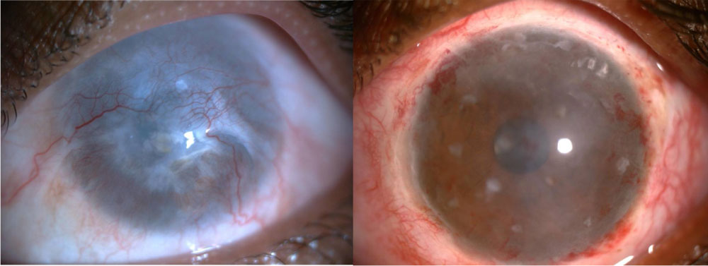
Pre-operative picture (Complete caption)Post-operative picture (at 3 months).
Leave a Comment