

Dr. Shreyas Temkar, S17031, Dr. Shorya Vardhan Azad, Dr.Rohan Chawla, Dr. Rajpal
Introduction
Vascular disorders form a major part of pediatric vitreoretinal disorders. Vascular disorders in children include conditions like retinopathy of prematurity (ROP), familial exudative vitreoretinopathy, Norrie disease, Incontinentia pigmenti, Coats disease and rare entities like vasculitis.[1] Most of these disorders present with peripheral retinal vascular abnormalities. Though clinical examination can provide sufficient clues to diagnosis, fluorescein angiography serves as an indispensible investigation to understand the pathogenesis, extent of disease and the need for treatment. Conventional techniques of fluorescein angiography are mostly non-contact procedures requiring patient cooperation. Also the field of view with most fundus cameras is limited, hence the documentation of peripheral fundus abnormalities is met with difficulty.[2] Various modalities of widefield imaging have been described in children,[3–6] but Retcam remains the preferred modality of imaging in pediatric patients. Retcam (Clarity Medical Systems, Inc.) is a widefield imaging device specifically developed for use in pediatric population. It is a contact type imaging system and hence can be used in small children during screening sessions (eg. ROP screening) or for fundus evaluation and documentation under anaesthesia.[7] The latest version, Retcam 3 has option for fluorescein angiography and has been shown to be useful tool for imaging vasculature in pediatric patients.[8–12] This study was conducted to assess the utility of Retcam fluorescein angiography in patients with pediatric vascular disorders.
Method
This was a retrospective study carried out in 43 eyes of 22 pediatric patients in whom conventional fundus imaging and fluorescein angiography was not possible or in whom additional procedures like laser or cryotherapy were planned as a part of management. The study was conducted at Dr Rajendra Prasad Centre for Ophthalmic Sciences, All India Institute of Medical Sciences. Patients were recruited from the retina and ROP services of the hospital. Written informed consent was taken for performing Retcam imaging, fluorescein angiography and expected interventions like laser or cryotherapy. Clearance from Pediatrician was obtained in all small babies and patients with systemic illness. Retcam imaging was carried out either under pediatrician monitored sedation (in babies < 3months) or under general anesthesia (in bigger children). Fundus imaging & fluroscein angiography was done using Retcam 130 degree wide field lens. Intravenous Sodium fluorescein (20%) was injected at dosage of 0.04 ml/kg or 6.6 mg/kg. Fluroscein angiography guided treatment (Laser /cryotherapy) was carried out wherever required. Follow up visits were planned based on disease type, severity and decision of the surgeon. Follow up imaging sessions were carried out wherever required to know the regression of the disease.
Results
Retcam guided imaging and FFA was carried out in 43 eyes of 22 patients.Patients included 7 females and 15 males.Patients age ranged from 6 weeks to 10 years.Diagnosis of patients included 5 cases of ROP, 5 cases of Coats disease, 4 cases of FEVR, 2 cases of ROP sequalae, 3 cases of congenital retinal folds, 1 case of double optic nerve head, 1 case of suspected vasculitis and 1 case of Incontinentia pigmenti.
Cases of ROP included 3 cases of aggressive posterior ROP (APROP) and 2 cases of vascular arrest following injection Anti VEGF for APROP.In 2 cases of APROP, fluorescein angiography was helpful in delineating the vascularized retina from avascular parts and to identify the avascular loops, which tend to get missed on routine clinical examination. Neovascularization of the iris and TVL could be clearly picked up with anterior focusing.In two cases of vascular arrest post injection clinical examination and color photography could not reveal the extent of retinal vascularization. However, FFA helped to clearly identify the junction of vascularized and avascular retina.
All 5 cases of Coats diseases were seen in males and were unilateral. FFA revealed telengectatic and aneurysmal changes and areas of avascular retina. (Figure 1a and 1b)
All cases of FEVR had bilateral disease.Out of 8 eyes, 3 had close funnel RD and rest of the 5 had stage 2 disease in 4 eyes and stage 1 disease in one eye.In two eyes retinal avascularity was seen in 360-degree peripheral retina, which is uncommon(usually avascularity is limited to temporal retina). (Figure 2a, 2b)
In two cases of ROP sequelae, 1 eye of each patient had a total retinal detachment.The other eye in first patient had avascular retina in Zone 3 and second patient had a free-floating venous loop causing vitreous hemorrhage. (Figure 3a, 3b)
One patient had clinical anophthalmos in one eye and evidence of double optic nerve head in the other eye. (Figure 4a, 4b) FFA clearely demonstrated evidence of 2 independent circulations arising from each of the optic nerve heads. One patient of suspected retinal vasculitis had both eye sclerosed vessels. Additionally FFA could show evidence of bilateral peripheral avascularity. FFA could demonstrate bilateral temporal retinal vascularity in one case of Incontinentia pigmenti.
Out of 3 cases of congenital retinal folds, two cases had family history of FEVR in either sibling or the parent.In one patient the cause of CRF could not be identified. (Figure 5a, 5b)
Complications and difficulties
None of the patients in the study developed anesthesia complications or allergic reactions. In 1 patient of APROP, who had extensive iris neovascularization, imaging of the late phases was met with difficulty due to leakage of the dye into the anterior chamber obscuring the fundus details.
Treatment
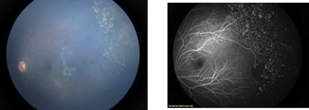
23 eyes received fluorescein angiography assisted laser treatment by Laser Indirect Ophthalmoscopy. Direct visualization of the FFA images on the Retcam screen helped the surgeon to precisely identify and treat the vascular abnormalities and avascular retina. (Figure 6) Two eyes received cryotherapy for the treatment of vascular abnormalities in Coats disease.4 eyes received retreatment with laser based on persistence of disease/avascular retina on FFA on follow-up. (Figure 7a, 7b)One eye received retreatment with cryotherapy. Regression of specific disease entities was seen in all eyes without any recurrence.
Discussion
The purpose of this study was to evaluate the utility of Retcam FFA in the management of pediatric retinal vascular disorders. In this study we intended to seeif FFA would provide more details compared only to clinical examination or imaging only. Retcam assisted FFA not only was able to match the clinical findings but also provided excellent details of the fundus pathology not appreciated on color photography. FFA in most of the diseases was able to pickup the extent of retinal avascularity, vascular abnormalities like telangiectasia, new vessels, avascular loops, free floating venous loop and so on.
Treatment in most of the adult vascular diseases is guided by fluorescein angiography. Since conventional FFA including the Spectralis and Optos require patient cooperation, Retcam serves as an excellent tool to image the pediatric patients not comfortable with the conventional imaging.
Retcam imaging has been shown to be an important tool in the management of retinopathy of prematurity. It has been extensively used for screening, documentation of disease, monitoring progression and telescreening purpose. The fluorescein angiography facility in Retcam has been used in ROP for diagnosis, to guide in treatment and to know the effectiveness of interventions. It has been shown to pickup subtle findings, which tend to be missed on indirect ophthalmoscopy or simple imaging.[8,9,13–15]The use of Retcam fluorescein angiography in other pediatric diseases has been reported less in the literature.[10–12]
In our retrospective study a wide range of pediatric retinal vascular disorders were included. In cases of APROP, FFA helped to identify the junction of vascularized retina and most importantly the avascular loops. Successful and early regression of disease depends on identification and treatment of all the avascular area including these loops. FFA helped in precisely identifying these pathologies and guided in treatment. In cases of aggressive posterior ROP, intravitreal injections of anti-VEGF have shown to be very effective in control of disease and aids in progression of vasculature. However in up to 20% of cases, vascular arrest can occur and clinically it would be difficult to decide further management. FFA in such cases would help to highlight the status of disease.[16–18] In both of our cases, FFA helped to delineate the area of vascularized retina but there was no evidence of any leak. Laser treatment was carried out in both the patients since the duration from injection was more than 6 weeks and hence no more vascularization progression was expected.
The application of Retcam FFA in the management of Coats disease has been extensively studied.[11,12] In all 5 patients, Retcam was able to pickup areas of capillary non-perfusion and telangiectactic abnormalities. FFA guided laser/cryo therapy was carried out in 4 patients and was extremely useful to identify subtle pathologies not picked up on clinical examination.
FEVR is a congenital retinal vascular abnormality characterized by exudation, retinal neovascularization and traction. It progresses in various stages and early treatment results in better visual and anatomical outcomes.[19] In 5 out of 8 eyes that had early disease, FFA helped to identify the extent of avascular retina and also vascular abnormalities. These eyes were successfully treated with laser. FFA was helpful in identification of persistent neovascularization and capillary non-perfusion in 2 eyes, which were not evident clinically. These eyes received laser retreatment with successful regression of disease.
Other few interesting pathologies were picked up in our study like – free floating venous loop causing vitreous hemorrhage, double optic nerve head etc.
There are a few limitations in the study. This was a retrospective review with variable duration of follow-up. We did not do a direct comparison between clinical examination based treatment and FFA guided treatment.However, FFA guided treatment provided insight into understanding of the specific disease pathologies. Since the surgeon can do real time FFA guided treatment, all pathologies can be efficientlydealt with better outcomes.
Conclusion
Retcam assisted FFA is an extremely useful investigation in the management of pediatric retinal vascular diseases. The widefield images captured help to document the subtle peripheral vascular pathologies that tend to be missed on clinical examination. Retcam FFA guided treatment results in excellent visual and anatomical outcomes.
References
- Leonard Feiner, Jonathan L Prenner. Retinal Vascular Disease in Children.Reviewof Ophthalmology. Published 12 April 2006.
- Nagiel A, Lalane RA, Sadda SR, Schwartz SD. ULTRA-WIDEFIELD FUNDUS IMAGING: A Review of Clinical Applications and Future Trends. Retina Phila Pa 2016;36(4):660–78.
- Magnusdottir V, Vehmeijer WB, Eliasdottir TS, Hardarson SH, Schalij-Delfos NE, Stefánsson E. Fundus imaging in newborn children with wide-field scanning laser ophthalmoscope. Acta Ophthalmol (Copenh) 2017;
- Kang KB, Wessel MM, Tong J, D’Amico DJ, Chan RVP. Ultra-widefield imaging for the management of pediatric retinal diseases. J Pediatr Ophthalmol Strabismus 2013;50(5):282–8.
- Fung THM, Yusuf IH, Xue K, Smith LM, Patel CK. Heidelberg Spectralis ultra-widefield fundus fluorescein angiography in infants. Am J Ophthalmol 2015;159(1):78-84.e1-2.
- Fung THM, Muqit MMK, Mordant DJ, Smith LM, Patel CK. Noncontact high-resolution ultra-wide-field oral fluorescein angiography in premature infants with retinopathy of prematurity. JAMA Ophthalmol 2014;132(1):108–10
- Wu C, Petersen RA, VanderVeen DK. RetCam imaging for retinopathy of prematurity screening. J AAPOS Off Publ Am Assoc Pediatr Ophthalmol Strabismus 2006;10(2):107–
- Azad R, Chandra P, Khan MA, Darswal A. Role of intravenous fluorescein angiography in early detection and regression of retinopathy of prematurity. J Pediatr Ophthalmol Strabismus 2008;45(1):36–9.
- Purcaro V, Velia P, Baldascino A, Antonio B, Papacci P, Patrizia P, et al. Fluorescein angiography and retinal vascular development in premature infants. J Matern-Fetal Neonatal Med Off J Eur Assoc Perinat Med Fed Asia Ocean Perinat Soc Int Soc Perinat Obstet 2012;25 Suppl 3:53–6.
- Escudero J, Borras F, Fernández MA, Domínguez C. [Fluorescein angiography with Retcam in incontinentia pigmenti: a case report]. Arch Soc Espanola Oftalmol 2009;84(10):529–32.
- Koozekanani DD, Connor TB, Wirostko WJ. RetCam II Fluorescein Angiography to Guide Treatment and Diagnosis of Coats Disease. Ophthalmic Surg Lasers Imaging Off J Int Soc Imaging Eye 2010;1–3.
- Suzani M, Moore AT. Intraoperative fluorescein angiography-guided treatment in children with early Coats’ disease. Ophthalmology 2015;122(6):1195–202.
- Zepeda-Romero LC, Oregon-Miranda AA, Lizarraga-Barrón DS, Gutiérrez-Camarena O, Meza-Anguiano A, Gutiérrez-Padilla JA. Early retinopathy of prematurity findings identified with fluorescein angiography. Graefes Arch Clin Exp Ophthalmol Albrecht Von Graefes Arch Klin Exp Ophthalmol 2013;251(9):2093–7.
- Blair MP, Shapiro MJ, Hartnett ME. Fluorescein angiography to estimate normal peripheral retinal nonperfusion in children. J AAPOS Off Publ Am Assoc Pediatr Ophthalmol Strabismus 2012;16(3):234–7.
- Guagliano R, Barillà D, Bertone C, Maffia A, Periti F, Spallone L, et al. Fluorescein angiography-based diagnosis for retinopathy of prematurity: expert-non expert comparison. Eur J Ophthalmol 2013;23(6):881–6.
- Lepore D, Quinn GE, Molle F, Baldascino A, Orazi L, Sammartino M, et al. Intravitreal bevacizumab versus laser treatment in type 1 retinopathy of prematurity: report on fluorescein angiographic findings. Ophthalmology 2014;121(11):2212–9.
- Lorenz B, Stieger K, Jäger M, Mais C, Stieger S, Andrassi-Darida M. RETINAL VASCULAR DEVELOPMENT WITH 0.312 MG INTRAVITREAL BEVACIZUMAB TO TREAT SEVERE POSTERIOR RETINOPATHY OF PREMATURITY: A Longitudinal Fluorescein Angiographic Study. Retina Phila Pa 2017;37(1):97–111.
- Tahija SG, Hersetyati R, Lam GC, Kusaka S, McMenamin PG. Fluorescein angiographic observations of peripheral retinal vessel growth in infants after intravitreal injection of bevacizumab as sole therapy for zone I and posterior zone II retinopathy of prematurity. Br J Ophthalmol 2014;98(4):507–12.
- Gilmour DF. Familial exudative vitreoretinopathy and related retinopathies. Eye Lond Engl 2015;29(1):1–14.
Figure legends
1a and 1b – Widefield color image showing extensive exudation in a case of Coats disease. Corresponding FFA showing aneurysmal changes and capillary non-perfusion areas not appreciated on color imaging.
2a and 2b – Case of FEVR showing temporal ridge. FFA clearly demonstrates profuse temporal leak.
3a and 3b – Free floating venous loop in a case of regressed ROP causing vitreous hemorrhage.
4a and 4b – Double optic nerve head in the fellow eye of a child with anophthalmos.
5a and 5b – Congenital retinal fold in a patient with family history of FEVR.
6 – Retcam FFA assisted laser in a case of FEVR.
7a and 7b–Followup images at 6 weeks of same patient as in figure 1a and 1b showing temporal avascularity and few telangiectasia. Localized laser augmentation was done.
Fig 1a and 1b
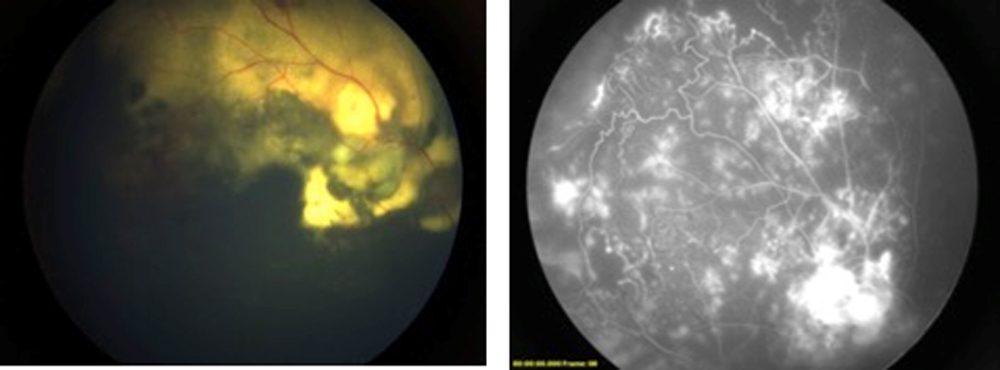
Fig 2a and 2b
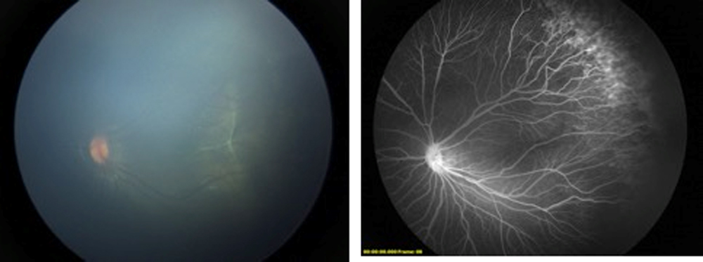
Fig 3a and 3b
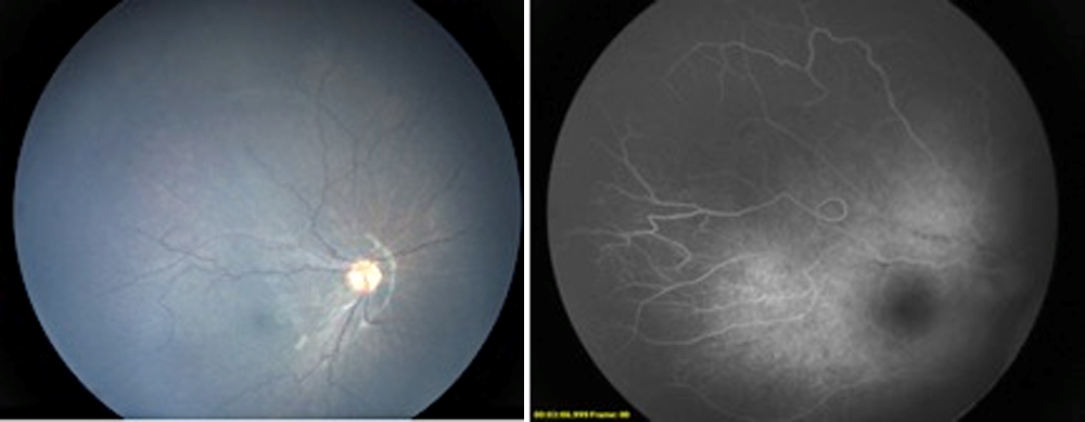
Fig 4a and 4b
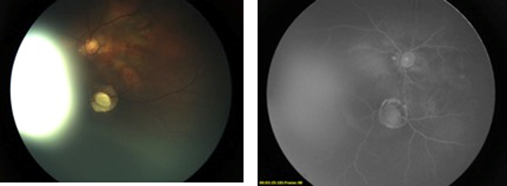
Fig 5a and 5b
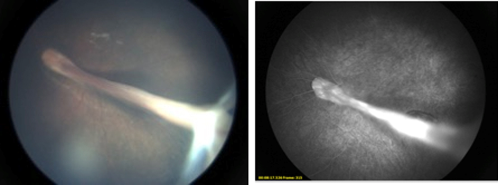
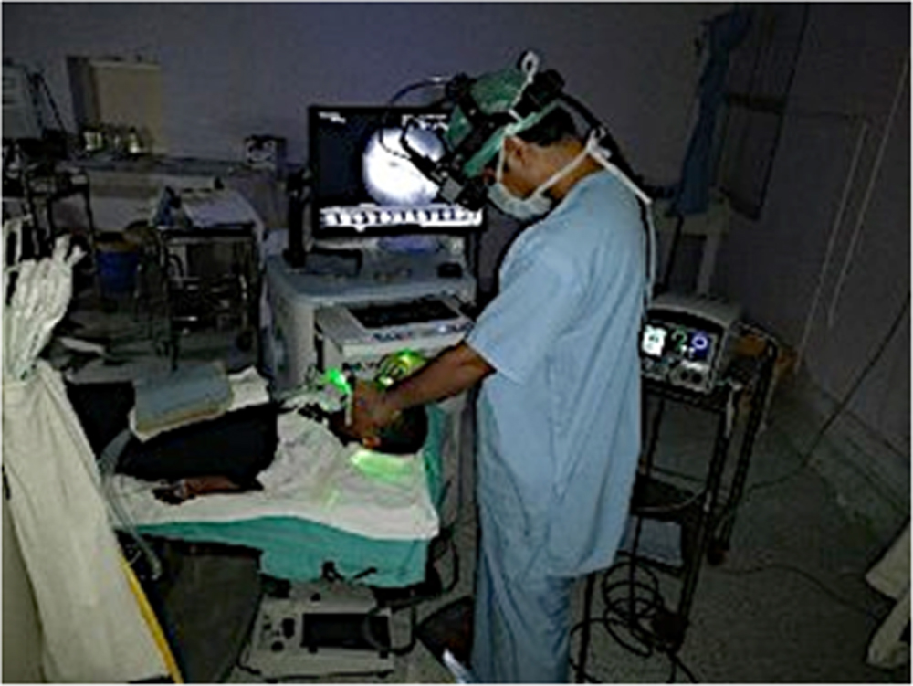
Fig 7a and 7b
Leave a Comment