
Dr. Sanjoy Chowdhury, C06721, Dr.priyanka, Dr. Sneha Kumari
Abstract:
Conjunctiva overlying pterygium (pterygial conjunctiva) is not at fault, hence this can be used as auto graft sparing other areas (Name: Conjunctival in situ auto graft: CISAG).Aim: To study surgical and morphological outcome of CISAG in pterygium. Methods: Pterygial conjunctiva is dissected from 1mm inside limbus, rest of the neck and apex were rrhexcised. Fibrotic tissues were meticulously dissected from conjunctiva and sclera. Curuncular end was placed along limbus over bare sclera. Clotted blood helped to keep graft in place. Weekly follow up for 1 month and monthly for a year. Apical pterygial conjunctiva was sent for HPE. Results: total 10 cases of primary pterygium, 2 recurrent pterygium and 2 cases of combined cataract and pterygium. No recurrence was observed Conclusion: no suture, no glue no other area traumatised. Our results were similar to other auto graft techniques but this new technique named CISAG saves time, tissue and recurrence
Surgical treatment of pterygium is complicated by its recurrence.Bare sclera has highest recurrence rate.To reduce this high incidence of recurrence covering the exposed sclera with grafts were introduced. These includes conjunctival auto grafts(CAG)with or without stem cells, amniotic membrane transplant (AMT).Different adjuvant therapy ranging from beta irradiation to antimitotic agents were also started. Thesewere not devoid of complications due to cytotoxicity hence lost theirpopularity. Pterygium itself has origins from cyto toxic effect of UV radiation. Moreover unstable tear film and cytotoxic effects of UV radiation can incite vasculoendothelial growth factors (VEGF) production which can give rise to pterygium formation.
Histopathologically conjunctiva is not at fault in pterygium. This is an elastotic degeneration of Subconjunctival tissues. So conjunctiva overlying pterygium can be used for covering bare sclera which can give stable tear film and protect from UV radiation thus preventing recurrence.
Aim of the study is to see the surgical and morphological outcome of a new technique “CONJUNCTIVAL IN-SITU AUTOGRAFT” (CISAG).
Methods:
Conjunctiva overlying the pterygium is dissected 1 mm inside the limbus after Subconjunctival injection of air. This helps in dissection and to examine the extent of sub conjunctival degeneration. Rest of the neck and apex is rrhexcised which lead s to relatively smooth scleral surface this will stabilise tear film posteoperatively.Conjunctiva is dissected free of all pterygium i.e. fibrotic Subconjunctival tissue. Curuncular end of the conjunctiva is placed along the limbus over the blood already clotting over bare sclera…CISAG is adhered by fibrin secreted from the clot below the graft.Followup was done weekly for 4 weeks ,this is the adhesion time and the futher monthly follow up for one year.Histopathological study of apical pterygial conjunctiva was done.
Results:
10 cases of primary pterygium, two recurrent pterygium and 2 case of CISAG done along with cataract surgery were included in this study No recurrence was observed. Withcataract surgery procedure becomes technically difficult CISAG may get displaced.
Discussions:
No suture, noglue, no other area is traumatised .No tissue wastage. Other methods of conjunctival auto grafts are also done with good results. Our results are similar but this newtechnique savestime, tissue and the eye from recurrence.Conjunctival pigmentations and presence of Stocker’s line are bad prognostic signs.
Conclusion:
CISAG is a new technique of pterygium surgery which is more physiological in nature as it does not traumatise other healthy portion of eyeball.
Limitations: It is a short study, not all cases were supported by histopathological examination.
References:
- Philipp WSpeicherLHumpel C. Expression of vascular endothelial growth factor and its receptors in inflamed and vascularized human corneas. Invest Ophthalmol Vis Sci 2000;412514- 2522
PubMed - Elizabeth Clearfield, Valliammai Muthappan, Xue Wang, and Irene C Kuo. Conjunctival autograft for pterygium.Published in final edited form as: Cochrane Database Syst Rev.; 2: CD011349. doi:10.1002/14651858.CD011349.pub2
Illustration:
Figure 1. Post op CISAG: Well-placed graft
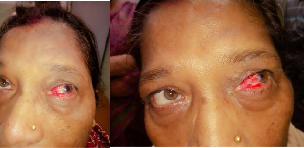
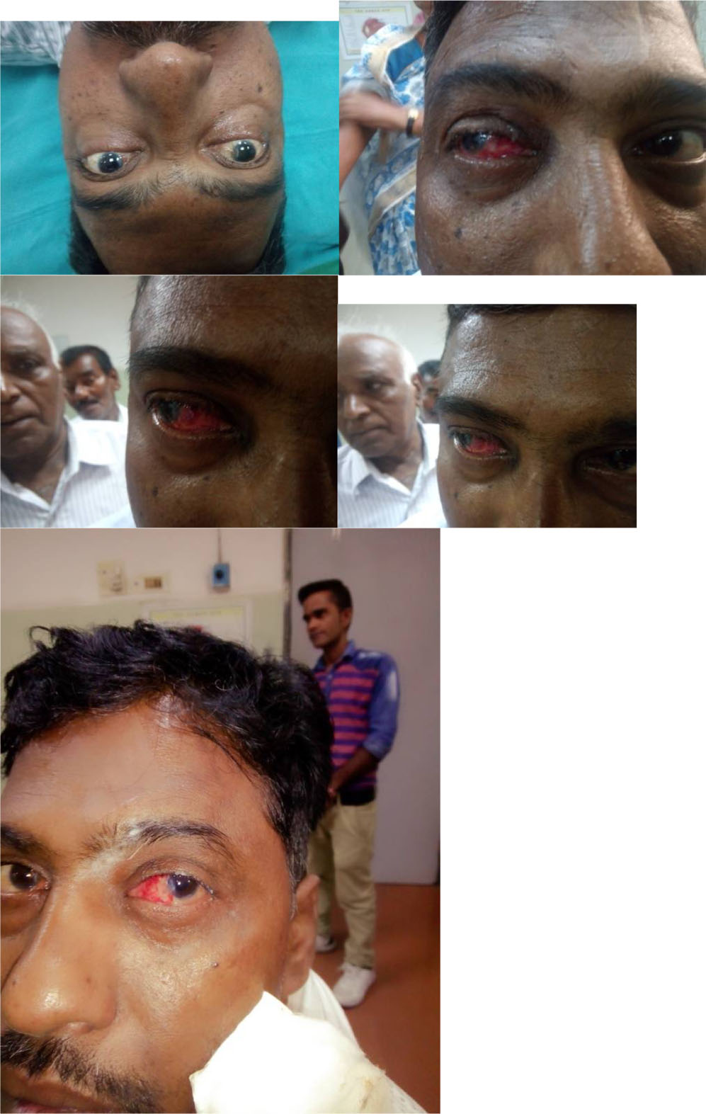
Figure 2.LE: 2months postop CISAG combined with SICS.RE: Pre and postop CISAG with SICS.
Leave a Comment