
Dr. Nishikant Borse, B10775
AIM:
To study the enhancing of the potential of brilliant blue G (BBG) for intraoperative staining of the internal limiting membrane (ILM) with respect to contrast using a Stain – Pick – Stain method
INTRODUCTION
Internal limiting membrane (ILM) peeling is one of the most challenging procedures in vitreous surgery. It is now routinely performed as a part of the surgical procedure for Macular Hole, Epiretinal Membrane surgery etc. This has been made easier with selective staining of this optically clear tissue with a number of vital dyes like brilliant blue G (BBG) and indocyaninegreen.BBG is the most commonly used dye for this purpose in India
Unfortunately the ILM does not show a good staining in many cases due persistent vitreous remnants or epiretinal membranes over the ILM. This can lead to a traumatic ILM dissection, retinal tears, retinal hemorrhage etc.
Commonly a re- staining of the ILM or staining the ILM after a fluid air exchange is used to enhance the ILM staining in such situations. However in most of the cases these measures may be of limited use.
We describe a stain-pick-stain method use intra operatively for enhancing the ILM staining potential of the BBG.
METHOD:
This was a prospective, interventional, non-comparative clinical case series.The authors analyzed 20 consecutive chromovitrectomy interventions in patients with macular holes, vitreomacular traction syndromes, or refractory macular edema. The patients with epiretinal membranes were excluded from the study as the epiretinal membranes are known to have a negative influence on BBG staining of the ILM.
All the patients underwent a complete ophthalmic clinical examination including best corrected visual acuity (BCVA), applanation intraocular pressure, Goldmann visual fields, color and red free photographs, and optical coherence tomography (OCT) (Cirrus OCT 500, Carl Zeiss MeditechInc, Jena, Germany) preoperatively, and every four weeks after surgery, for a period of 20 weeks.
All patients in the study underwent either a 23/25 GuageVitrectomy.
All cases were surgically naïve eyes without and previous vitreous / retinal procedures. After completing the vitrectomy the internal limiting membrane was stained with BBG Solution 0.05 % w/v (Ocublue Plus / Aurolab, India) followed by picking the ILM with an ILM forceps. After the initial pick the ILM was re stained in 10 eyes.
In the other 10 eyes the ILM was just re stained without a initial pick.
For each stain, the BBG solution was injected slowly over the macular area with a blunt tipped cannula and washed out after about 30 seconds.Theoperating surgeon evaluated the contrast subjectively.
ILM staining was noted intraoperatively as good, faint or absent.
The ease of dissection etc was not considered as these may be influenced by factors other than the staining modality.
RESULTS:
Objective contrast improved in all cases with the stain – pick – stain method as compared with a simple re-staining of the ILM with BBG.
All the eyes had a good stain in the Stain – Pick – Stain Method group as compared to 4 eyes in the restained eyes group.About 6 eyes in the re-stained group had a faint staining.The Stain-Pick-Staining of theILM was good in all cases and allowed for an easy and atraumatic removal of the ILM.
There was no evidence of any retinal pigment epithelium abnormality or nerve fiber layer defect seen in the red-free photographs during any of the postoperative visits of all patients. No ocular or systemic side effects were noted.
DISCUSSION:
The vitreal side of the ILM has epiretinal cellular or fibrocellular proliferation and vitreal remnants as compared to the retinal side.
Studies have shown that the epiretinal cellular or fibrocellular proliferation on the vitreal side of the ILM was higher (58%) as compared to the retinal side of the ILM (23%).*
Transmission electron micrographs of specimens removed by brilliant blue G assisted internal limiting membrane (ILM) peeling in macular hole surgery showed an undulated retinal side without retinal debris or with small fragments of Muller cells. The vitreal side of the ILM shows cellular proliferation in five of nine eyes. #
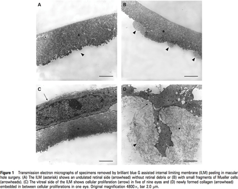
Thus the vitreal side of the ILM has many cellular or fibrocellular components that can interfere with the uniform staining of the ILM with BBG.
The initial pick exposes the retinal side with very minimal fibrocellular elements to the BBG stain, enabling a uniform dark staining of the retinal side of the ILM. This well stained flap has a better contrast than the vitreal side of ILM.
It enables the easy grasping of this flap and hence assists in a good ILM peeling.It is especially useful in myopic eyes where the contrast is very less.
The limitations of this study are the objective nature of evaluating the contrast introducing a possibility of the surgeon’s bias & the very small size of this study.
Leave a Comment