
Dr. Vaibhav Khanna, Dr. Zain Khatib
INTRODUCTION
Smartphone ophthalmoscopy has been in use since many years now, and in today’s world a number of smartphone adapters are available at a reasonable price for capturing fundus images.With the ever-growing Internet and Mobile industry in India, it is important for us to realize the impact of smartphones and their capability in capturing good quality fundus images. A normal fundus camera is still out of reach in many ophthalmic setups; thus creating a huge gap for fundus imaging and documentation, which can be easily filled by using such mobile adapters.
One such adapter has been developed by our team, which allows the use of any smartphone to capture fundus images, edit them with the help of free software’s and then share them using social media.The traditional smartphone ophthalmoscopy includes holding a 20D/28D lens in one hand and a smartphone with an always-on flashlight and camera in the other hand. Due to instability of this system, the images were found to be out of focus and had a long learning curve to master this technique. (Figure 1)
Here we have evolved the technique of smartphone indirect ophthalmoscopy by making an adapter, which can be stabilized on the slit lamp for capturing fundus images.
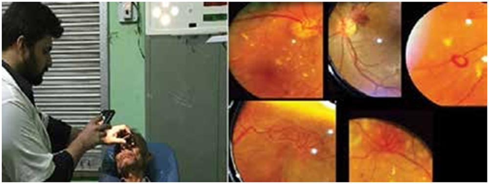
Figure 1: left: Traditional smartphone ophthalmoscopy; Right: Images obtained
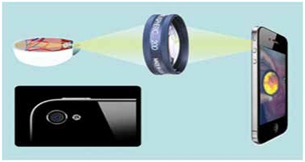
Figure 2- Principle of working
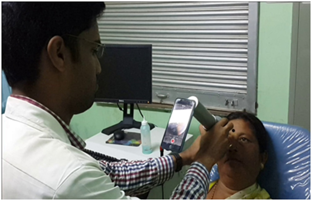
Figure 3- Handheld adapter
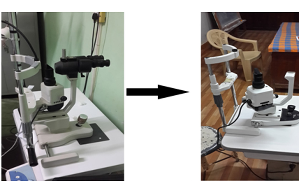
Figure 4- Left- Old slit lamp; Right- Slit lamp after removing observation part
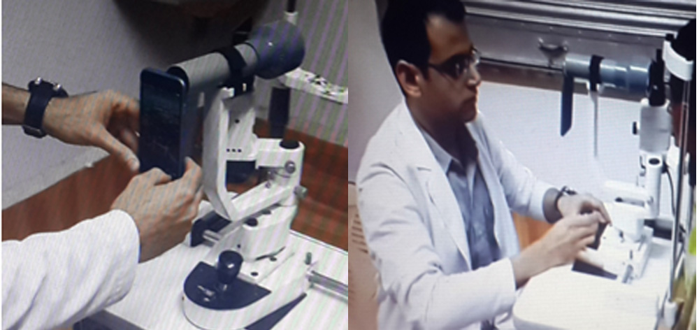
Figure 5- Left- Mounting a handheld adapter on the slit lamp
Right- Our first Slit Lamp smartphone adapter in use.
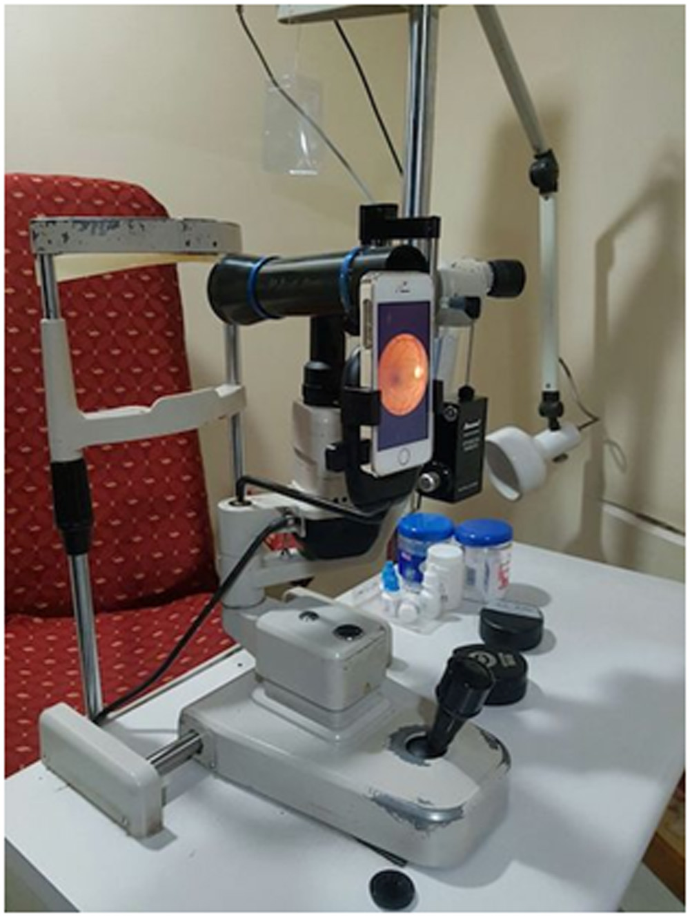
Figure 6- Slit lamp mounted smartphone adapter
MATERIALS AND METHODS
The basic material required for smartphone fundus imaging is an adapter which are easily available or it can be made on your own also [6], a 20D/28D lens and a smartphone with a camera above 5 megapixel preferably.
Principal of working: The LED light of the phone acts as a coaxial light source to illuminate the patient’s retina, and is turned ‘ON’ throughout the procedure. This system works as an indirect ophthalmoscope wherein the camera creates a digital image of the fundus on the phone screen through the condensing lens (Figure 2)
The traditional method of smartphone ophthalmoscopy is quite difficult to master since it involves everything to be done handheld; with smartphone camera, the 20D/28D lens and well dialated patient eye all to be in one single line. The distance of the 20D/28D lens from the eye and the distance between the smartphone and the lens have to be fixed by the examiner’s hand in order to have a well-focused image of the fundus. Because of such difficulties this method was not popular and soon was replaced by many smartphone adapters.
The smartphone adapters help keeping the smartphone camera and the lens in line and at a constant distance from each other (Figure 3). The image quality did improve with the handheld adapters to a certain level, but it had a long learning curve since this system was unstable, as holding the adapter with a phone at a constant distance from the patient’s eye with one handwas a difficult task. (Figure 3)
This handheld design was inspired from a blog written by DrBijuRaju[7](Figure 3)In order to improve further on the stability of the adapter, it was placed over a slit lamp with the help of PVC pipes after trying many permutation and combinations. (Figure 4,5) This adapter was now stabilized on the universal hole present in all slit lamps at the base of the arm, which was earlier used for placing the Hruby lens.(Figure 6)
Details of the procedure are as follows:
- Good pupillary dilatation prior to procedure
- LED Flashlight: Remain “on” throughout the procedure
- Camera: Video record mode, Manual focus
- Saving, editing of images using any simple Android/iOS based software
- Sharing of images through social networking media
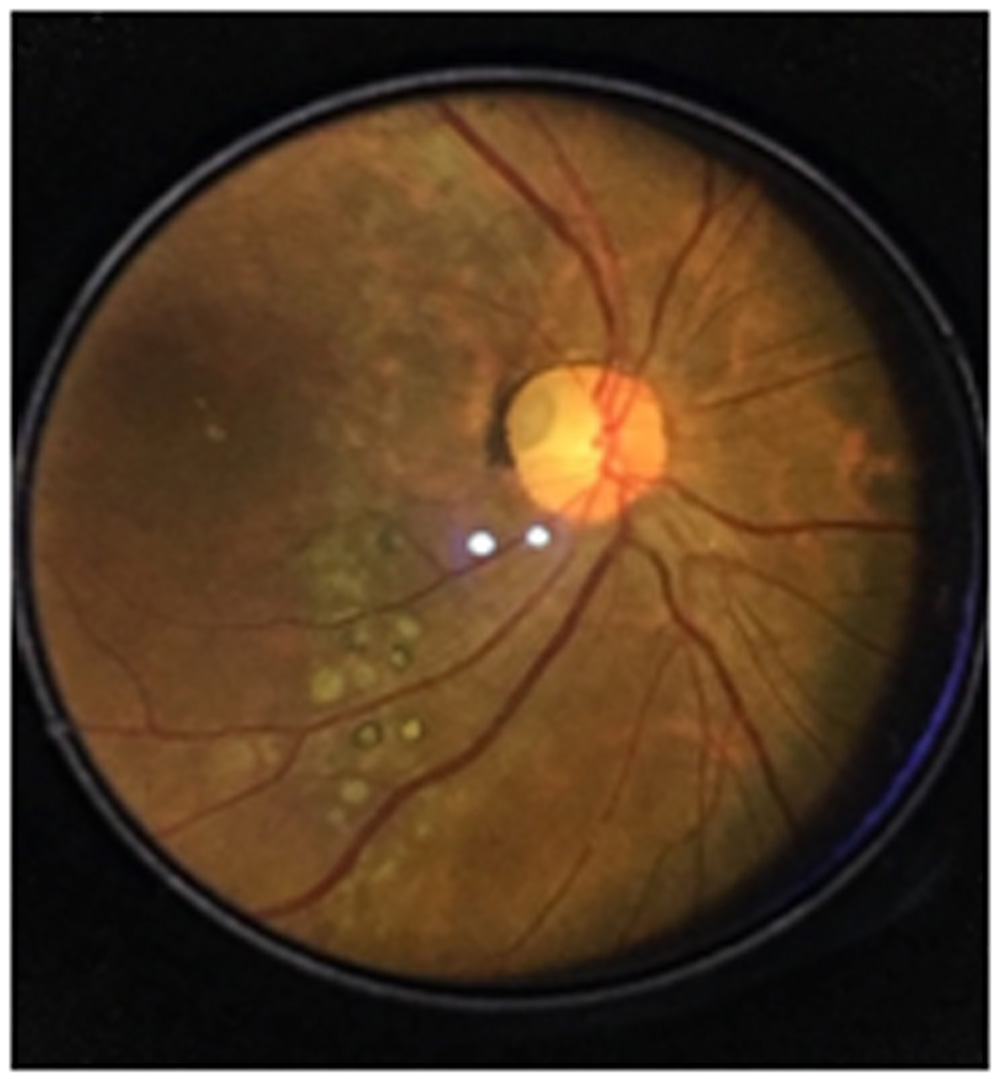
Figure 7- Optic disc pit with ILM folds and retinal atrophic patches
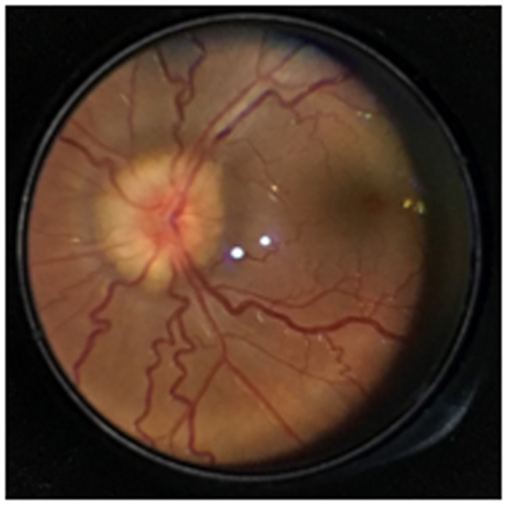
Figure 8- Papilledema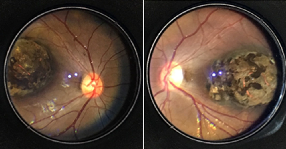
Figure 9- Macular Dystrophy
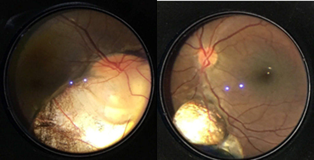
Figure 10- Typical Coloboma
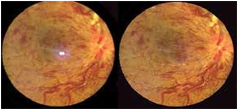
Figure 11- CRAO left image- with central reflection, Right image – without central reflection
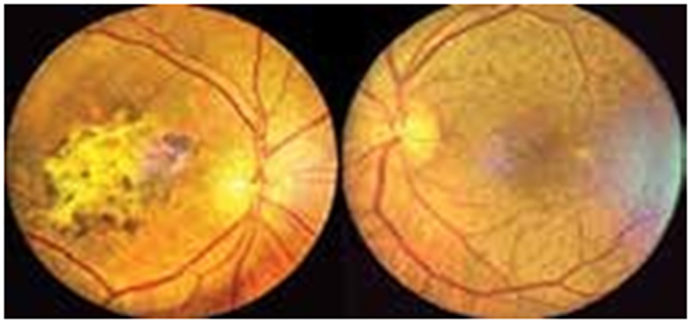
Figure 12- ARMD, Left – Wet, Right- Dry
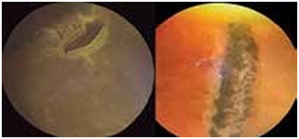
Figure 13- Retina Periphery, Left- Horse shoe tear, Right – Lattice degeneration
RESULTS
With the slit lamp mounted adapter, we were able to obtain excellent quality fundus image (Figure 7-13). The adapter has been made for any kind of smartphone. Since the adapter is placed on the slit lamp it is highly stable and has a shorter learning curve as compared to the traditional smartphone indirect ophthalmoscopy and other handheld adapters. The slit lamp adapter allows the examiner to do fine adjustments in order to focus the fundus image.
The only drawback that we noticed was a central reflection artifact that was seen due to reflections from the lens surface. However this reflection can be edited in the phone itself using readily available software free of cost at android/iOS platform.
With the use of 20D lens we were able to capture 30 degrees of retinal image of the posterior pole. For viewing peripheral part of retina i.e. beyond equator the same adapter was used as a handheld device. Also by taking two images in the same examination at a different angle we can obtain stereoscopic effect, which gives a good depth perception by using Google Cardboard hardware as an attachment to our phone.
TABLE 1
| FUNDUS CAMERA | MOBiRET CAM | |
| Image quality | Good | Good |
| Central Reflection | Absent | Present, can be eliminated with the help of software editing |
| Image orientation | Erect | Inverted |
| FFA | Can be done | Not possible |
| Cost | Costly | Extremely cost effective |
DISCUSSION
Smartphone ophthalmoscopy has been in practice since many years now; it has evolved from the traditional method i.e. without any adapter to the slit lamp mounted adapter.It serves as a good teaching tool for demonstrating various ocular fundus pathologies. Since it is easy to operate and has a short learning curve it can be used even by residents, nursing staff students to document various pathologies.
Moreover, most commercially available fundus cameras are out of the reach of many practitioners and institutions due to their high cost. Our smartphone adapter proves to be an extremely cost effective alternative to these imaging devices, retaining the same image quality. The differences between our instrument and the commercial fundus camera’s available are summarized in Table 1. Being light and portable, the instrument can be easily carried from one place to another and can be mounted on all types of slit lamps. It can also be used in peripheral centres where slit lamps are not available as a hand held instrument for various screening programmes (Diabetic retinopathy screening, ROP screening etc.).
Finally, the role of smart phones in telemedicine cannot be overemphasized. The use of smart phones for capturing fundus images serves as an extremely efficient way to share and transfer images instantly via social networking media (Whatsapp, Facebook. etc.)
CONCLUSION
By making a simple and cost effective fundus camera adapter, we could maximize the potential of smart phones to capture high quality fundus images which are comparable to those obtained from commercially available fundus cameras.
REFERENCES
- Dyaberi R, Bajantri YB, Khatib ZI. Smartphone indirect ophthalmoscopy: For screening evaluation, and documentation of the ocular fundus. J Vis Sci2015;1:13-6.
- BijuRaju, Raju N S D. “Regarding fundus imaging with a mobile phone: A review of techniques”. Indian Journal of Ophthalmology, 2015 February. pp. 170- 172.
- Luis J. Haddock, David Y. Kim, Shizio Mukai, “Simple, Inexpensive Technique for High Quality Smartphone Fundus Photography in Human and Animal Eyes”. Journal of Ophthalmology 2013, Volume 2013, Article ID 518479.
- Mahesh P Shanmugam, Mishra Divyansh KC, Madhukumar R, Rajesh Ramanjalu, Shrinivasulu Y Reddy, Gladys Rodrigues, “Fundus imaging with a mobile phone: a review of techniques”. Indian Journal of Ophthalmology, 2014 September pp. 960-962.
- Bastawrous, “Smartphone fundoscopy”. Ophthalmology 2012;119:432.-3.
- Raju, B., Raju, N.S.D., Akkara, J., Pathengay, A., 2016. Do it yourself smartphone fundus camera – DIYretCAM. Indian Journal of Ophthalmology 64, 663. doi:10.4103/0301-4738.194325
- BijuRaju. “DIY retCAM”. Vitreoretinalsurgeon.blogspot.com, May 2015
Leave a Comment