
Dr. Amrita Sawhney, S17593, Dr. Harsh Rathod, Dr. Shaloo Bageja, Dr. Ashok Kumar Grover
Purpose : To study the wide spectrum of diagnosis in fifty cases of lacrimal gland disorders along with their epidemiological, radiological, laboratory and histological data.
The recognition of the newer entities like IgG4 and Rosai Dorfman disease have dramatically altered the spectrum of histopathologically diagnosed lacrimal gland lesions.
Rosai-Dorfman disease, also known as sinus histiocytosis with massive lymphadenopathy (SHML) is a rare disorder of unknown aetiology characterized by nonmalignant proliferation of distinctive histiocytes within lymph node sinuses and other extranodal sites. About 43% of patients have extranodal manifestation in the eye, upper respiratory tract, salivary gland, skin, bone, meninges and CNS1. The reported ophthalmic manifestations include eyelid and orbital mass, and rarely, uveitis 2. Management options include surgical excision or debulking for lesions in surgically accessible locations, and systemic corticosteroids, chemotherapy or radiotherapy. Massive or recurrent orbital disease or significant residual lesion following surgical debulking may be treated with systemic corticosteroids or chemotherapy. Chemotherapy has also been used to relieve sight threatening optic nerve compression 3.
Immunoglobulin G4-related disease is a relatively newly defined emerging entity which is characterized by mass forming lesion, lymphoplasmacytic infiltration rich in IgG4 positive plasma cells and raised IgG4 levels in 60-70 % cases. 4 It was first identified in a group of patients with autoimmune pancreatitis with raised serum IgG4 levels5 and was recognized as a systemic condition in 2003.6
IgG4 –RD can involve several organs of the body including orbit, lymph node, skin, lungs, salivary glands, thyroid, aorta, biliary tract, breast, kidney, retroperitoneum , CNS etc. Orbit is the most common extrapancreatic site to be involved by this disease entity.7 The most frequently involved site in IgG4 –related orbital disease is the lacrimal gland (IgG4 –related dacryoadenitis). Other sites in the orbit that can be involved are – extraocular muscles (IgG4 –related orbital myositis), orbital soft tissues (IgG4 related orbital inflammation), eyelids and orbital nerves. Orbital IgG4 –related disease is predominantly seen in adults with no gender predilection.8
Materials and method
It was a retrospective study of 50 cases with histopathologically diagnosed lacrimal gland lesions from October 2002 to May 2017. Their epidemiological profile (including age and sex), presenting complaints, laboratory results (like CRP, ESR, serum ACE, serum IgG4 etc.), orbital imaging and histological findings were recorded.
Results
We diagnosed 50 cases of lacrimal gland disorders histopathologically from 2002-2017.
The average age at presentation in our studywas 41 years(12-71years) with male:female ratio of 1:1.3 (22 males and 28 females).
The lesions comprised of IgG4 related orbital disease ( 7cases) (Figure 1) , Rosai Dorfman disease (5cases) (Figure 2), infective lesions like tubercular dacryoadenitis (4cases) , bacterial dacryoadenitis (2 cases) and viral dacryoadenitis (3 cases), non specific inflammation (12 cases) and sarcoidosis (1 case). Commonly occurring tumour were pleomorphic adenoma (7 cases) ,adenoid cystic carcinoma (4 cases), lymphoma (4 cases) and myoepithelioma (1case).
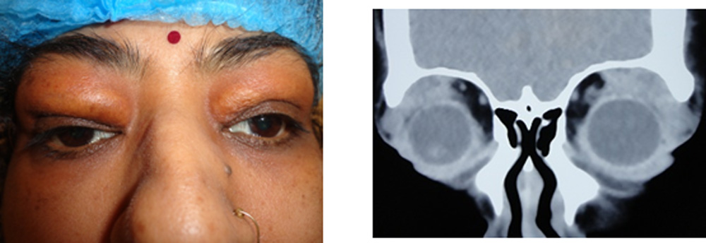
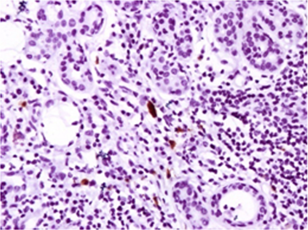
Figure 1: a) 55 year old female presenting with bilateral upper lid swelling (Bilateral dacryoadenitis) was diagnosed as IgG4 related orbital disease. b) CTscan showing soft tissue swelling in superotemporal quadrants on both sides. c) Histopathological diagnosis of IgG4 lacrimal gland disease was based on IgG4 positive plasma cells with fibrosis (IHC under 40X showing IgG4 positive plasma cells)
c
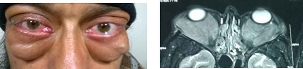
Figure 2 :
- 35 year old male presented with bilateral gradually progressive proptosis
- Soft tissue mass in intra and extra-conal compartment, hypo-intense on T1 and T2 W images with encasement of the optic nerve in intra-conal space
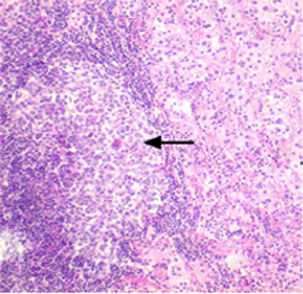
- Histopathology showing fibroadipose tissue with histiocytes. Histiocytes showing evidence of emperipolesis
Discussion
Better awareness of the newer entities like IgG4 disease and Rosai Dorfman disease have dramatically altered the spectrum of histopathologically diagnosed lacrimal gland lesions.
The clinical correlation of this unusual spectrum of diagnosis offers a great insight into the lacrimal gland disorders.
References
- Foucar E, Rosai J, Dorfman RF: The ophthalmologic manifestation of sinus histiocytosis with massive lymphadenopathy.Am J Ophthalmol 1979, 87:354-367.
- Vemuganti GK, Sekhar GC, Indira K: Multifocal Rosai-Dorfman disease of periorbital tissues spanning 15 Years – a case report.Orbit 2001, 20:297-300.
- Lee-Wing M, Oryschak A, Attariwala G, Ashenhurst M: Rosai-Dorfman disease presenting as bilateral lacrimal gland enlargement.Am J Ophthalmol 2001, 131:677-678.
- Stone JH, Zen Y, Deshpande V. IgG4-related diseasestyle=”font-size: . N Engl J Med. 2012; 366:539-551.
- Hamano H, Kawa S, Horiuchi A ,et al. High serum IgG concentrations in patients with sclerosing pancreatitis. N Engl J Med. 2001;344:732-738.
- Stone JH. IgG4- related disease:nomenclature,clinical features, and treatment. Semin Diagn Pathol. 2012;29:177-190.
- Divatia M, Kim SA, Ro JY. IgG4 –related sclerosing disease, an emerging entity: a review of a multi-system disease. Yonsei Med J. 2012;53:15-34.
- Kubota T, Moritani S, Katayama M, et al. Ocualr adnexal IgG4-related lymphoplasmacytic infiltrative disorder. Arch Ophthalmol. 2010;128:577-584.
Leave a Comment