

Dr. Girish Bharat Velis, V16197, Dr. Annamalai O, Dr. Sahil Bhandari, Dr. S Bala Murugan
Introduction
Choroidal folds are the undulations involving the inner choroid, Bruch’s membrane, the retinal pigment epithelium and sometimes the overlying retina. Eversince Nettleship in 1884, first described them in a patient with atrophic papilloedema, they have generated curiosity among clinicians.1 Multiple etiologies have been associated with them, but still they are often projected as idiopathic without proper work up for underlying etiology either due to lack of awareness or inadequate resources. Literature review revealed, idiopathic choroidal folds to be commoner in the past, however recently disease specific choroidal folds are commonly reported. Trivial findings such as these should not be ignored as they may be the only sign of some serious underlying pathology.
Through this article, we describe the various ocular co-morbidities associated with choroidal folds in atertiary eye care center in South India and want to highlight the basic necessary work up to rule out associated pathologies before labelling them as idiopathic.
Materials and methods
Medical case records of patients diagnosed with choriodal folds at Vitreoretina services of Aravind Eye Hospital Pondicherry, from 1st Jan 2013 to 31st Dec 2015 were retrieved. The data obtained from the case records included demographic details, presenting complaints with duration, uncorrected and best corrected visual acuity (BCVA) in snellen, refraction, axial length, intraocular pressure (IOP) by non contact tonometry, anterior and posterior segment findings, diagnosis and treatment details. Colour vision and central fields were assessed in patients with optic disc edema. Ultrasound B Scan was done in patients suspected to have posterior scleritis. At follow-up, change in BCVA, resolution of the primary pathology causing the choroidal folds and disappearance of the choroidal folds if any were noted. Colour vision, central fields and Ultrasound B Scan were repeated, if required, to assess the progress. Institutional Review Board (IRB) at Aravind Eye Hospital, Pondicherry had approved this study. This research adhered to the tenets of the Declaration of Helsinki.
Statistical analysis
Frequency (percentage) was used for categorical variables. Chi-square test or Fisher’s exact test was used to assess the difference between two categorical variables. P-value <0.05 was considered statistically significant. All statistical analysis was done by statistical software STATA 11.1 (StataCorp, College Station, TX).
Results
A total of 169 eyes of 121 patients with a male to female ratio of 1:2 were included into the study. Mean age was 42.2 ± 13 (Range= 13 – 65) years. Mean follow up was 24.8 ± 38 months.No difference was noted between laterality of eyes involved. Painless defective vision (76/169, 45% eyes) and asymptomatic routine eye evaluation (65/169, 38% eyes) were the two most common presenting scenarios in patients diagnosed with choroidal folds. Other less common chief complaints were pain (3 eyes), headache (1 eye), trauma (3 eyes) and painful defective vision (7 eyes). Fourteen eyes were diagnosed to have choroidal folds on day 1 of post cataract surgery. Duration of complaints (median=1.5 months) was available in 87 eyes only. Mean duration was 12 ± 32 months. Etiological analysis revealed idiopathic CF in 136 eyes (80.5%) and disease specific or non idiopathic CF in 33eyes (19.5%) as mentioned in Table 1 and Pie chart.
Table 1 Etoilogical analysis
| Diagnosis | Frequncy(%) |
| Idiopathic | 136(80.5) |
| Posterior Scleritis | 7(4.1) |
| Posterior Uveitis | 2(1.2) |
| VKH | 6(3.6) |
| SO | 1(0.6) |
| Post OP Hypotony | 1(0.6) |
| Post Filtering Surgery | 8(4.7) |
| Disc Related | 5(3.0) |
| Rare | 3(1.8) |
| Total | 169(100) |
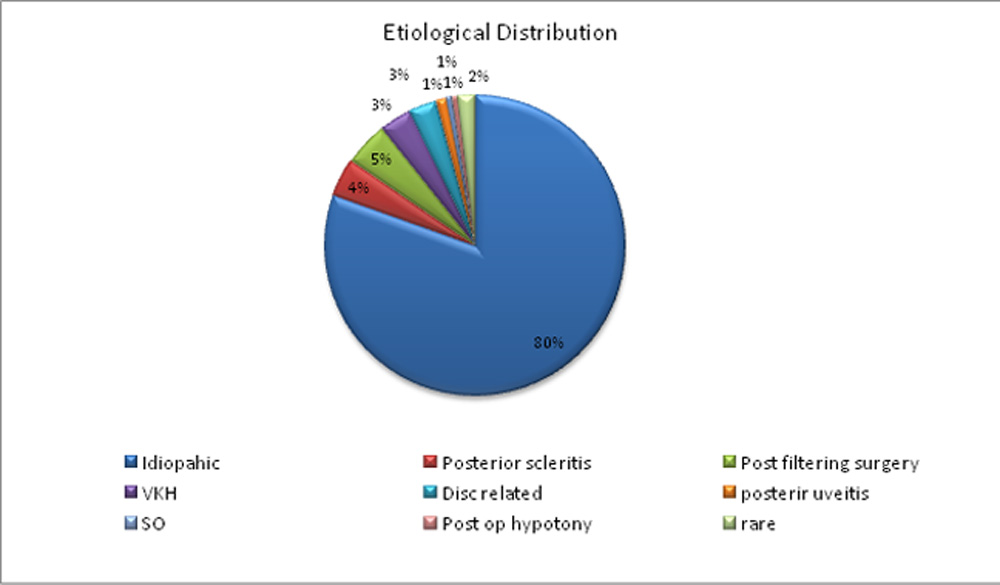
An overall significant difference (p=0.001) was observed between mean best corrected visual acuity (BCVA) at presentation (0.21) and last follow up (0.12) but in subgroup analysis of eyes diagnosed with non-idiopathic choroidal folds (CF’s) no such difference was observed (p=0.15). Between group (idiopathic versus non-idiopathic CF’s) analysis of BCVA at presentation and last follow up, revealed a significantly better BCVA (p=0.01) in eyes with idiopathic CF’s. Mean intraocular pressure (IOP) was 14.6 ± 5.21 mmHg (R= 2-35mmHg). A comparative analysis of demographic variables between idiopathic CF’s and non-idiopathic CF’s is mentioned in Table 2.
Table 2: Comparative analysis of demographic variables between idiopathic and non-idiopathic CF’s
| Continuous variables | Idiopathic CF’s (Mean±SD) | Non-Idiopathic CF’s (Mean±SD) | P-value |
| UCVA | 0.57±0.36 | 0.60±0.35 | 0.63 |
| Presenting BCVA | 0.18±0.30 | 0.33±0.35 | 0.01 |
| Axial length | 21.8±1.0 | 22.7±0.95 | 0.02 |
| Duration of complaints (months) | 15.2±35.7 | 1.05±2.7 | 0.08 |
| Intraocular pressure (mmHg) | 14.9±4.6 | 13.2±6.6 | 0.08 |
| Follow up (months) | 32.3±42.7 | 3.8±7.0 | 0.002 |
| Follow up BCVA | 0.09±0.17 | 0.21±0.23 | 0.01 |
Details of refraction, associated ocular co-morbidities, associated anterior and posterior segment findings and etiological diagnosis has been mentioned in Table 3 and Table 4.
Table 3: Details of refraction, associated anterior segment findings and ocular co-morbidities in eyes diagnosed with choroidal folds
| Refraction | No. of eyes (n) | Anterior segment findings | No. of eyes (n) | Ocular co-morbidities | No. of eye (n) |
| Simple hypermetropia | 34 | Nil | 148 | Nil | 149 |
| Simple hypermetropic astigmatism | 2 | Ocular surface inflammation | 2 | POAG | 6 |
| Compound hypermetropic astigmatism | 2 | Anterior segment inflammation | 11 | PACS/PACG | 9 |
| Not available | 131 | Post surgical hypotony | 7 | Secondary OAG | 4 |
| corneal perforation | 1 | Secondary ACG | 1 |
Table 4: Details of associated posterior segment findings and etiological diagnosis in eyes diagnosed with choroidal folds.
| Posterior segment findings | No. of eyes (n) | Etiological diagnosis | No. of eyes (n) |
| Nil | 131 | Idiopathic | 136 |
| Small disc | 1 | Posterior Scleritis | 7 |
| Central serous retinopathy | 3 | Posterior Uveitis | 2 |
| CNVM/PCV | 4 | VKH | 6 |
| Posterior scleritis | 7 | SO | 1 |
| Disc edema | 3 | Post cataract Hypotony | 1 |
| NPDR | 4 | Post filtering surgery | 8 |
| Serous choroidal detachment | 4 | Disc related | 5 |
| CRVO | 1 | Miscellaneous | 3 |
| BRVO | 2 | ||
| Ischemic optic neuropathy | 2 | ||
| VKH | 6 | ||
| SO | 1 |
Axial length was documented for 64 eyes (38%) with a mean of 21.9±1.04mm. Only 3 eyes had axial length ≤20mm. Rest all 61 eyes were between the range of >20mm to <24mm. A detailed gender and laterality wise distribution of axial length is mentioned in Table 5.
Table 5: Gender and laterality wise distribution of axial length
| Gender | ≤20mm | >20 to ≤21mm | >21 to ≤22mm | >22 to ≤23mm | >23mm |
| Right eye | |||||
| Male | 1 | 3 | 4 | ||
| Female | 1 | 2 | 11 | 8 | |
| Left eye | |||||
| Male | 1 | 1 | 1 | 6 | |
| Female | 1 | 2 | 12 | 9 | 1 |
| Total | 3 | 7 | 25 | 24 | 15 |
Etiological diagnosis was categorized into two groups: idiopathic and non-idiopathic. Chi-square test was used to analyze the relationship between etiological diagnosis and gender, refraction, axial length, chief complaints, duration of complaints (≤6;>6 to ≤12; >12 months), presence or absence of anterior segment, posterior segment & ocular co-morbidites and presence or absence of B-scan pathology. All were found to have significant relation (p<0.05) except gender and axial length. Binary logistic regression analysis was thus performed taking into account the same group of variables. Gender was the only variable to give statistically insignificant results. Odds ratio of all other variables along with P-value has been mentioned in Table 6. Multivariate logistic regression analysis model using the above mentioned variables was not successful because of collinearity and absence of convergence.
Table 6: Binary logistic regression analysis for Etiological diagnosis (Non-idiopathic versus idiopathic CF’s)
| Independent Categorical Variable | Odd’s Ratio | P-value | Independent Categorical Variable | Odd’s Ratio | P-value |
| 1. Gender | 0.89 | 0.8 | 2. Refraction | 1.6 | 0.03 |
| 3. Axial length | 2.8 | 0.03 | 4. Chief complaints | 1.6 | 0.000 |
| 5. Duration of complaints | 0.06 | 0.01 | 6. Anterior segment co-morbidity | 91 | 0.000 |
| 7. Posterior segment co-morbidity | 15 | 0.000 | 8. Other Ocular co-morbidity | 3.3 | 0.02 |
| 9. B-scan pathology | 13.5 | 0.000 |
In the disease specific group, CF’s persisted in 5 eyes (3 in posterior scleritis and 2 in rare association) and resolved in 7 eyes (5 post filtering surgery, 1 posterior scleritis and 1 disc related) till the last follow up in the documented case records (Table 7).
Table 7 Frequency table for CF status at follow up in the non idiopathic group
| CF status at follow up | Diagnosis | Total
n(%) |
|||||||
| Posterior Scleritis
n(%) |
Posterior Uveitis
n(%) |
VKH
n(%) |
SO
n(%) |
Post OP Hypotony
n(%) |
Post Filtering Surgery
n(%) |
Disc Related
n(%) |
Rare
n(%) |
||
| Resolved | 1
(14.3) |
– | – | – | – | 5
(71.4) |
1
(14.3) |
– | 7
(100) |
| Persistent | 3
(60.0) |
– | – | – | – | – | – | 2
(40.0) |
5
(100) |
| Status not Documented | 2
(20.0) |
2
(20.0) |
2
(20.0) |
1
(10.0) |
1
(10.0) |
– | 1
(10.0) |
1
(10.0) |
10
(100) |
| Total | 6
(27.3) |
2
(9.1) |
2
(9.1) |
1
(4.6) |
1
(4.6) |
5
(22.7) |
2
(9.1) |
3
(13.6) |
22
(100) |
Discussion
The various causes of chroidal folds described include hypermetropic eyes with short axial length, optic disc edema, optic disc drusen, central serous retinopathy, choroidal tumours and effusion, conditions associated with proptosis such as retrobulbar masses, orbital cellulitis, thyroid related ophthalmopathy, inflammation such as posterior scleritis, post-surgical like after scleral buckling or vitrectomy, hypotony, in astronauts following long-duration space flight or idiopathic.2-7
Our study predominantly involved idiopathic CF compared to disease specific variety. In the non idiopathic group, CF were most common post filtering surgery (8 eyes) followed by posterior scleritis (7 eyes). Other causes consisted of Vogt Koyanagi syndrome (VKH), disc related, posterior uveitis, sympathetic ophthalmia (SO), post operative hypotony and rare associations like optic nerve dural ectasia, lacrimal gland tumor, adenoid cystic carcinoma

Fig 1 Fundus image showing linear CF in a case of adenoid cystic carcinoma along with the B scan image showing intraconal mass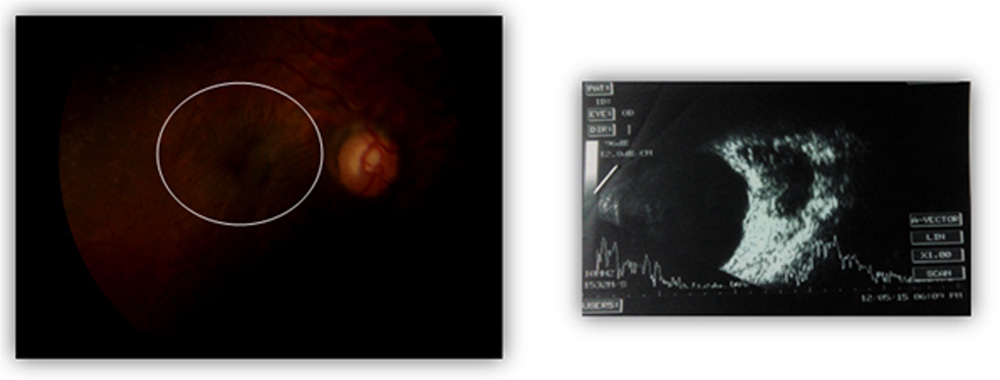
Fig 2 Fundus image showing radial CF in a case of optic nerve dural ectasia along with B scan image depicting the pathology
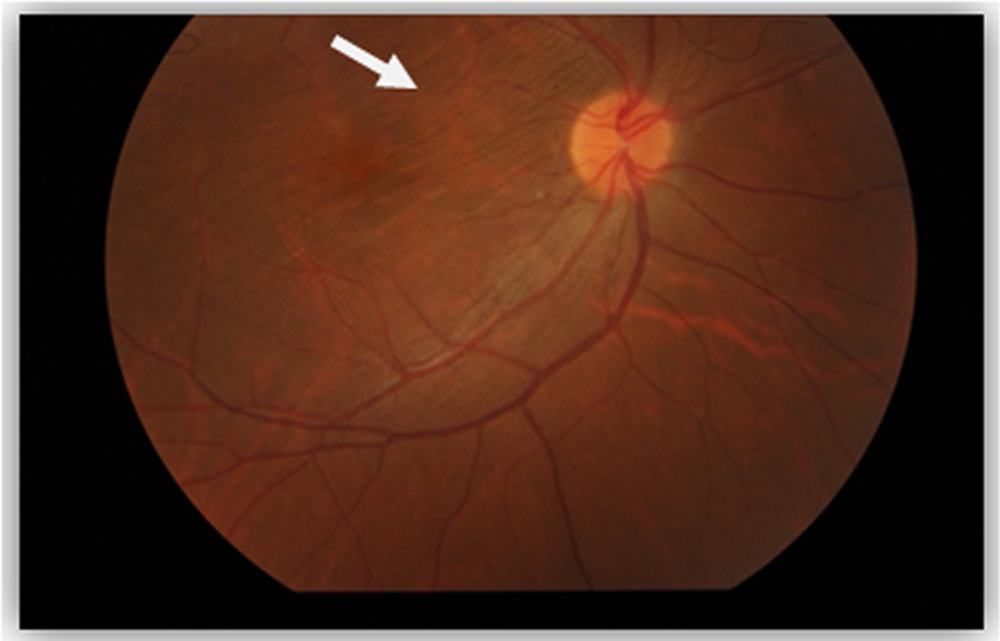
Fig 3 Fundus image showing linear CF in a case of lacrimal gland tumor above ON corresponding to the mass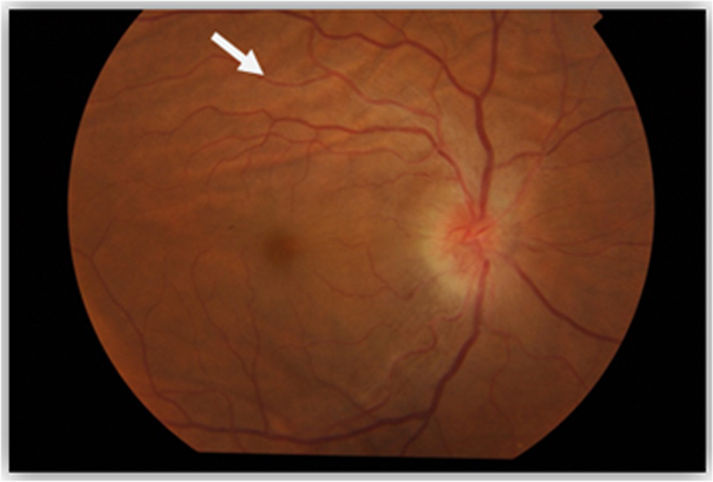
Fig 4 Fundus image showing CF in a case of AION
Our study highlights the importance of detailed work up of CF before judging them as idiopathic. As documented in the present study, the extensive causes of CF require case specific work up, complete ocular and systemic examination. It consists of visual acuity, perimetry, tonometry,fundus fluorescein angiography, ultrasonography – A scan and B scan, orbital CT scan and interdisciplinary examination (ear, nose, throat and neurological). Choroidal folds have been described in literature as an indicator of previous episode of posterior scleritis8, early sign of idiopathic intracranial hypertension9-11, indicator of previous inflammation of the orbit or paranasal sinuses12, h/o autoimmune disease or undiagnosed ocular inflammation.13
The present study has certain limitations in that being retrospective in nature, inconsistent follow up visits and lack of recent choroidal imaging (EDI). Because of the referral pattern of our hospital, we may have missed orbital and other described etiologies of the CF.
To summarise, CF is an important clinical sign with potential underlying causes and need proper detailed analysis.
References
- Nettleship E. Peculiar lines in the choroid in a case of post papillitic atrophy. Trans Ophthalmol Soc UK 1884;4:167–8
- Cangemi FE, Trempe CL, Walsh JB. Choroidal folds. Am J Ophthalmol. 1978 Sep;86(3):380-7.
- Bullock JD, Egbert PR. The origin of choroidal folds a clinical, histopathological, and experimental study.Doc Ophthalmol. 1974 Nov;37(2):261-93.
- Mader TH, Gibson CR, Otto CA, Sargsyan AE, Miller NR, Subramanian PS, Hart SF, Lipsky W, Patel NB, Lee AG. Persistent Asymmetric Optic Disc Swelling After Long-Duration Space Flight: Implications for Pathogenesis. J Neuroophthalmol. 2016 Dec 5.
- Poon JS, Vahdani K, Booth AP Chorioretinal folds in a patient with multiple myeloma treated with stem cell transplant. Eur J Ophthalmol. 2017 Mar 10;27(2):e46-e49.
- Liu DT, Chan CK, Fan DS, Lam SW, Lam DS, Chan WM. Choroidal folds after 25 gauge transconjunctival sutureless vitrectomy. Eye (Lond). 2005 Jul;19(7):825-7.
- Jaworski A, Wolffsohn JS, Napper GA. Aetiology and management of choroidal folds. Clin Exp Optom. 1999 Sep-Oct;82(5):169-176.
- Friberg TR. The etiology of choroidal folds. A biomechanical explanation. Graefes Arch Clin Exp Ophthalmol 1989;227(5):459–464
- Taban M, Kosmorsky GS, Singh AD, Sears JE. Choroidal folds secondary to parasellar meningioma. Eye 2007;21(1):147–150.
- Sharma M, Volpe NJ, Patel T, Kimmel A. Intracranial hypertension associated with acquired hyperopia and choroidal folds. Retina 1999;19(3):260–262
- Griebel SR, Kosmorsky GS. Choroidal folds associated with increased intracranial pressure. Am J Ophthalmol 2000;129(4):513–516
- Hyvarinen L, Walsh FB. Benign chorioretinal folds. Amer J Ophthalmol 1970; 70: 14-17
- Olsen TW, Palejwala NV, Lee LB, Bergstrom CS, Yeh S.Chorioretinal folds: associated disorders and a related maculopathy. Am J Ophthalmol 2014;157(5):1038–1047.e1
Leave a Comment