
Dr. Megha Gulati, G18578, Dr. Santosh Kumar Thakur, Dr. Rakhi Kusumesh, Dr.
Bibhuti Prassan Sinha
ABSTRACT : DTI is a technique that identifies white matter tracts (WMT) non-invasively using diffusion measurements. WMT are not visible with classical MRI or intra-operatively with microscope. DTI allows for accurate identification of patients who will develop visual deficits, and who therefore require more urgent surgical intervention and will help neurosurgeons to prevent destruction of the visual pathway while removing lesions adjacent to this WMT.
KEYWORDS : conventional MRI, MRI Tractography, Diffusion tensor imaging (DTI), visual pathway, optic chiasm
INTRODUCTION
Tumors and other lesions affecting the visual pathway may displace the fibres or infiltrate them, both having entirely different visual prognosis
Routine Magnetic Resonance (MR) sequences can evaluate the location of intracranial neoplasm. But the location relationship of neoplasm and white matter (WM) tracts nearby, the integrity of WM tracts and its infiltration by tumor tissues are not definite. The presence of T2 hypersensitivity in the optic nerves has been correlated with the degree of chiasm compression and the degree of visual impairement
Diffusion tensor tractography (DTT) is a computational procedure that reconstructs major fiber bundles in three-dimensional spaces based on their anisotropy properties. It is the most visual method for presenting target tracts from DTI data. It helps to determine the infiltration of white matter tracts by tumor, and provides evidence of degeneration of white matter tracts distal to tumor sites
CASE DESCRIPTION
CASE 1 : 46 years female with diminution of vision both eyes, left more than right and left eye disc pallor
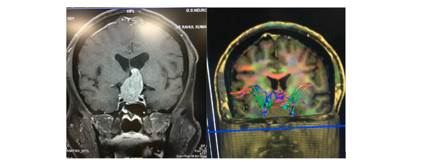
Figure 1 : Conventional MRI (left) showing large homogenous mass in sella with suprasellar extension and MRI tractography(right) showing splayed fibres of optic pathway around the lesion
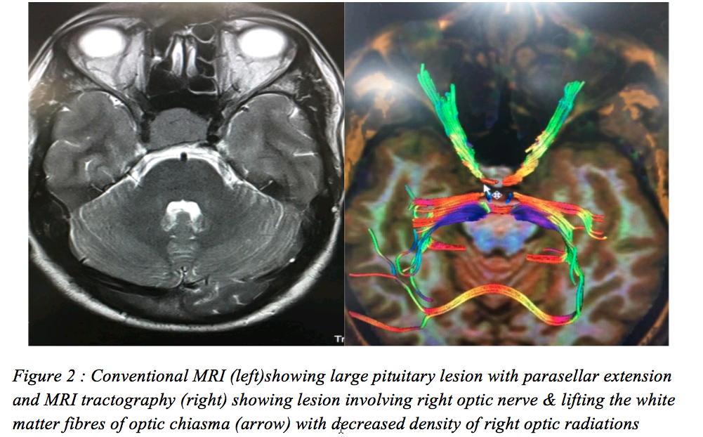
CASE 2 : 26 years male with diminution of vision right eye and temporal disc pallor; with clinically normal left eye
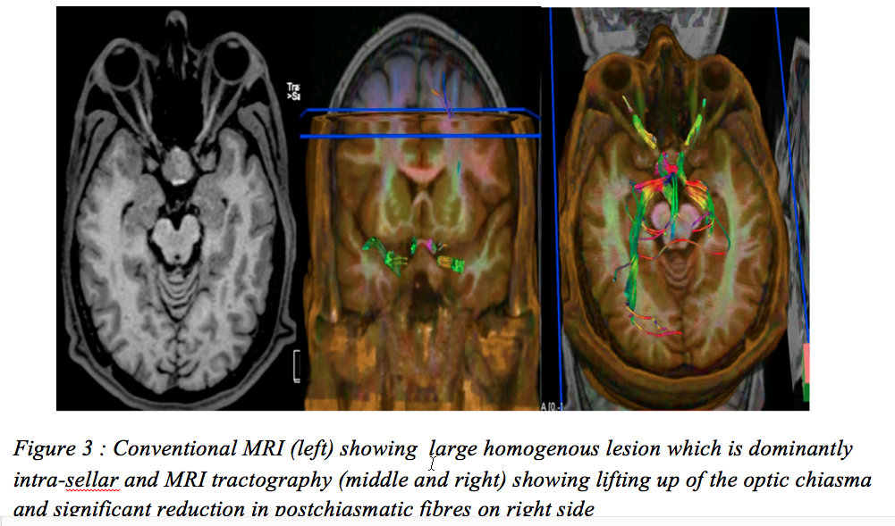
CASE 3 : Pituitary Lesion with dominant intra-sellar component. In such situations, the optic chiasma is lifted up and is pushed posteriorly and superiorly
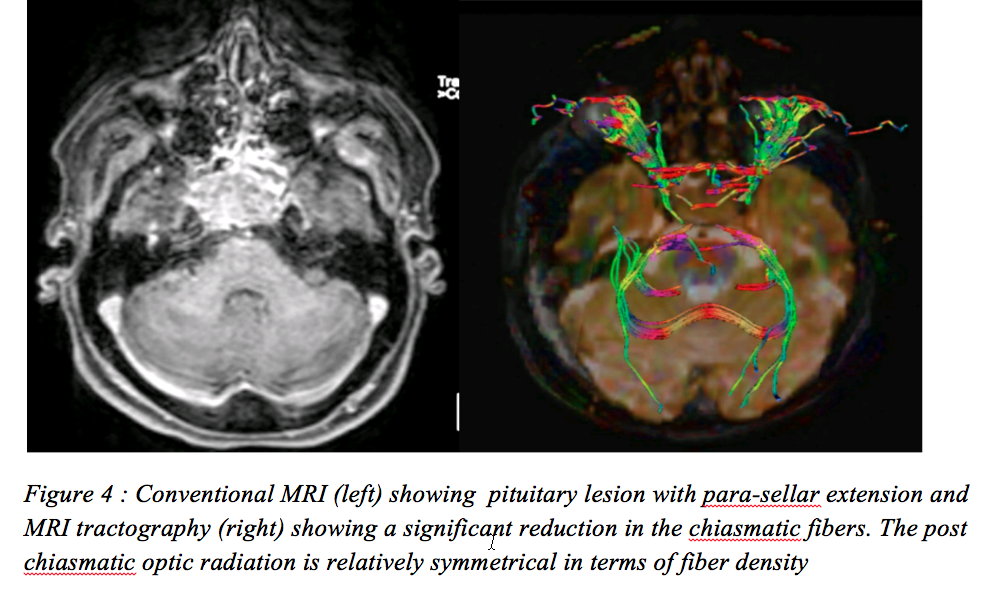
CASE 4 : Pituitary Lesion with dominant para-sellar component. In such situations, the optic chiasma is infiltrated and the fibers are interspersed with the tumor.
DISCUSSION
Diffusion tensor MR imaging (DT-MRI or DTI) techniques sample water motion in atleast 6 non collinear directions, providing information about both the rate and direction of water proton motion. DTI can noninvasively evaluate the WM integrity and fiber connectivity in vivo1 and thus important in disease states that disrupt fiber tract structure.
The parameters of DTI are called diffusion indices. Diffusion indices include the fractional anisotropy (FA) and mean diffusivity (MD). The FA value reflects the direction of and the MD value reflects the speed of diffusion, mainly.2
- FA reflects the directionality of molecular displacement by diffusion and vary between 0 (isotropic diffusion) and 1 (infinite anisotropic diffusion).
- MD reflects the average magnitude of molecular displacement by diffusion. The more the MD value, the more isotropic is the medium
These values can be expressed either as a number or as a directionally encoded colour map. A change in these indices provides information on underlying microanatomic changes or pathological changes of WM fiber bundles.
Diffusion tensor tractography (DTT) is a computational procedure that reconstructs major fiber bundles in three-dimensional spaces based on their anisotropy properties. It is the most visual method for presenting target tracts from DTI data. It helps to determine the infiltration of white matter tracts by tumor, and provides evidence of degeneration of white matter tracts distal to tumor sites (wallerian degeneration). DTT is been used for visualisation of tumor location relative to eloquent WM tracts and has been found to be beneficial in the neurosurgical planning and postoperative assessment
WM tracts may become oedematous, displaced, disrupted or infiltrated with neoplasms : 3
- Displaced, the WM tracts have abnormal pattern and location but normal fractional anisotropy
- Disrupted, the WM tracts will disappear in FA images with significant decrease in fractional anisotropy
- Infiltrated, the WM tracts had abnormal pattern and location. Its fractional anisotropy is decreased. But in FA images, the tracts still can be identified
Orbital tumor impresses the optic nerve and causes visual field changes. However, it is not clear whether impressed nerve is degenerated or necrotic. Now we can make judgment with MR-DTI whether the degeneration is reversible or not after years. The smaller the FA value is, the bigger possibility of necorsis, vice versa. This is because hydrones lose the direction dominant after tissue necorsis, and the anisotropy decreases and the FA value decreases, as a result, the isotropy and the MD value also increases.4
CONCLUSION
MRI Tractography over conventional MRI has the ability to reliably correlate with visual function in setting of optic chiasma compression and post-chiasmatic density of fibres and thus allows for accurate identification of patients who will develop visual deficits, and who therefore require more urgent surgical intervention ; as well as an intraoperative guide to surgery.
REFERENCES
- Bammer R, Acar B, Moseley ME. In vivoMR tractography using diffusion imaging. Eur J Radiol. 2003;45(3):223–234
- Mori S, vanZijl PC. Fiber tracking: principles and strategies-a technical review. NMR Biomde. 2002;15(7):468–480
- Ciccarelli O, Toosy AT, Parker GJ, Wheeler-Kingshott CA, Barker GJ, Miller DH, Thompson AJ. Diffusion tractography based group mapping of major white-matter pathways in the human brain. 2003;19(4):1545–1555
- Tropine A, Vucurevic G, Delani P, Boor S, Hopf N, Bohl J, Stoeter P. Contribution of diffusion tensor imaging to delineation of gliomas and glioblastomas. J Magn Reson Imaging. 2004;20(6):905–912.
Leave a Comment