
Dr. Sanjay Kumar Dhar, D12867, Dr. Archana Singh, Dr. Gaurav Kapoor
Abstract-
Purpose-
To study RNFL thickness and other ONH parameters in Amblyopic and better fellow eyes.
Materials and methods-
A total of 10 patients (05 patients with meridonial amblyopia,02 patients with Anisometropic amblyopia, 02 patients with strabismic amblyopia and 01 patient Isohypermetropic amblyopia) diagnosed as amblyopic were divided into two groups, Group A (Amblyopic eyes – 10 eyes) and Group B (Better fellow eyes – 10 eyes). All the eyes underwent visual acuity assessment, refraction (under cyclopegia), anterior segment evaluation and fundus evaluation. All the patients underwent ONH evaluation by Spectral-Domain optical coherence tomography. The ONH parameters studied were average RNFL thickness, rim area, disc area and cup volume. The data was analysed by paired t-test and Wilcoxon rank test.
Results-
The mean and standard deviation for RNFL thickness was 79.9 mm2 and 20.82 mm2 in group A and 82.8 mm2 and 17.61 mm2 in group B respectively. There was a trend of decreased RNFL thickness in Amblyopic eyes, although same was not statistically significant (P-value-0.19). None of the parameters studiedshowed statistically significant difference.
Conclusion-
There was a trend of decreased RNFL thickness in amblyopic eyes as compared to normal fellow eyes,although it was not found to be statistically significant.
Keywords– Amblyopia,Retinal Nerve Fibre Layer (RNFL) thickness,Optic Nerve Head (ONH).
Introduction-
Amblyopia is a disorder of decreased visual acuity and contrast sensitivity1. Previous studies have shown retinal changes in amblyopic eyes2, 3, however this was refuted later by other studies 4,5. Studies in the recent past with the help of SD-OCT, which has better speed and resolution, have reported variable results on retinal thickness in amblyopic eyes6, 7. Hence it is still a subject of exploration and further study.
In our study we have compared the RNFL thickness, disc area, rim area and cup volume between the amblyopic and normal fellow eye.
Material and Methods-
This prospective observational study was carried out at a tertiary care centre. All diagnosed patients of unilateral amblyopia were included in the study.A total of 10 patients (5 patients with meridonial amblyopia, 2 patients with Anisometropic amblyopia, 2 patients with strabismic amblyopia and 1 patient Isohypermetropic amblyopia) diagnosed as amblyopic were divided into two groups, Group A (Amblyopic eyes – 10 eyes) and Group B (Better fellow eyes – 10 eyes). All the eyes underwent visual acuity assessment, refraction (under cycloplegia), anterior segment evaluation and fundus evaluation. All the patients underwent SD-OCT for RNFL thickness (peripapillary), disc area, rim area and cup volume. The mean BCVA in amblyopic eyes was +0.64 logMAR and+0.22 logMAR in better fellow eyes before amblyopia treatment and +0.43 logMAR in amblyopic eyes and +0.14 logMAR in better fellow eyes after amblyopia treatment.
Statistical analysis was done using paired t-test and Wilcoxon rank test.
Results-
The study included 8 males and 2 females. The youngest patient was 6-years old and eldest was 15-years old. There were 5 cases (50%) of Meridonial amblyopia, 2 (20%) cases of Anisometropic amblyopia,02 (20%)cases of strabismic amblyopia and 1 (10%) case of Isohypermetropic amblyopia. The mean and standard deviation for RNFL thickness was 79.9 mm2 and 20.82 mm2 in group A and 82.8 mm2 and 17.61 mm2 in group B respectively. There was a trend of decreased RNFL thickness in Amblyopic eyes, although it was not statistically significant (P-value-0.19). Mean Rim area in group A was 1.74 mm2 and in group B was 1.57 mm2. (P value-0.31). Mean Disc area in group A was 2.57 mm2 and in group B was 2.41 mm2 .(P value-0.48). Mean Cup volume in group A was 0.1014 mm3 and in group B was 0.98 mm3. (P value-0.95). None of the parameters studied showed statistically significant difference.
| Parameters | T-test | Wilcoxon rank sign test | ||
| Mean | Difference | p-value | p-value | |
| Rim Area GroupA | 1.7 | 0.17 | 0.3 | 0.3 |
| Rim Area GroupB | 1.5 | |||
| Disc Area GroupA | 2.6 | 0.15 | 0.5 | 0.3 |
| Disc Area GroupB | 2.4 | |||
| CupVolGroupA | 0.1 | 0.004 | 0.9 | 0.5 |
| CupVolGroupB | 0.09 | |||
| RNFL Group A | 79.9 | -2.9 | 0.2 | 0.17 |
| RNFL Group B | 82.8 | |||
| Group APre Occlusion VisionLog MAR | 0.6 | 0.2 | 0.005 | 0.03 |
| Group APost Occlusion Vision Log MAR | 0.4 | |||
| Group B Pre Occlusion Vision Log MAR | 0.22 | 0.08 | 0.02 | 0.06 |
| Group B Post Occlusion Vision Log MAR | 0.14 | |||
*Highlighted = statistically significant i.e. p-value <0.05
Table 1. Mean difference in various parameters between Group A and B
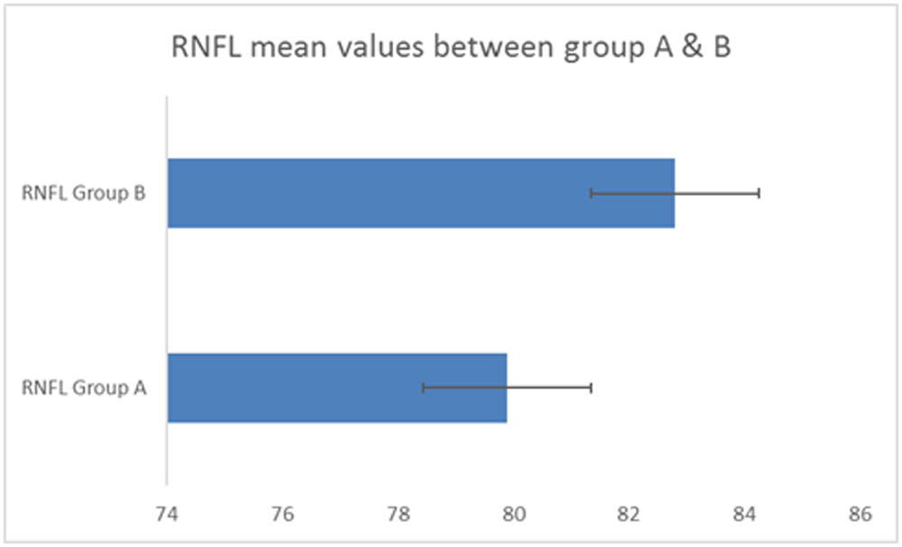
Graph 1. Mean difference in RNFL thickness between Group A and B
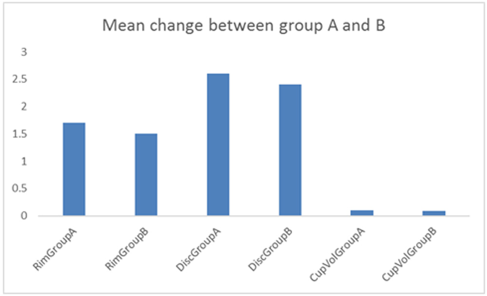
Graph 2. Mean difference in ONH parameters between Group A and B
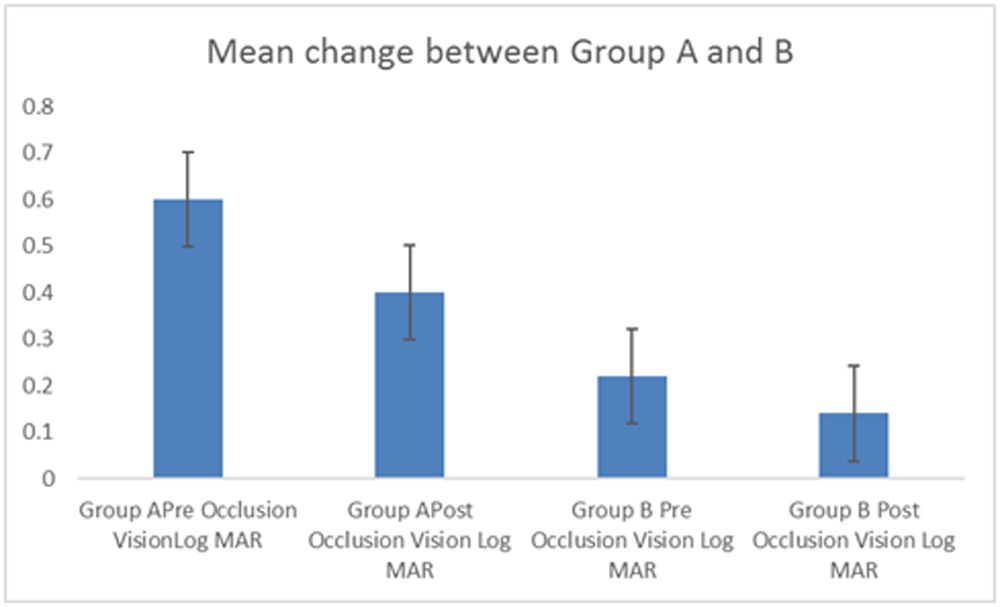
Graph 3. Mean difference in visual acuity (log MAR) between Group A & B
Discussion-
This study was done to find the difference between RNFL thickness and other ONH parameters (disc area, rim area and cup volume) between Group A (amblyopic eyes) and Group B (normal fellow eyes). A trend of decreased RNFL thickness in amblyopic eyes, which was different from Syunsuke Anaki et al.8 and Wu et al. 9our study results are also different from Al Haddad et al. 6 who observed no difference in the RNFL thickness in two groups. The limitation in our study was a small size and no comparison group of normal patient eyes.
The large number of evaluation done for amblyopic retinal thickness show variable results.There was a trend noted in amblyopic eyes of decreased RNFL thickness, same was not statistically significant.There was no statistical difference between Rim Area,Disc Area and Cup volume in the two groups.Our study results are different from the results of Al Haddad et al (RNFL thickness same) and opposite to the results of Wu SQ et al (RNFL thickness more in amblyopic eyes).The drawbacks of the study is small sample size and no normal group.
Conclusion-
The RNFL thickness was less in amblyopic eyes as compared to normal fellow eyes. Rimarea, Disc area and Cup volume did not show any statistically significant difference in two groups. The study results of our study should be seen after giving consideration to the small sample size , which can change the statistical significance if the sample size is increased. No consensus has reached so far regarding changes in the various ONH parameters in amblyopic eyes , hence there is a requirement of a larger study to evaluate the same .
References-
- Von Noorden GK. Amblyopia: a multidisciplinary approach. Proctor lecture. Invest Ophthalmol Vis Sci. 1985;26(12):1704–1716.
- McNeil NL. Patterns of visual defects in children. Br Ophthalmol. 1955;39(11):688–701.
- Ikeda H, Tremain KE. Amblyopia occurs in retinal ganglion cells in cats reared with convergent squint without alternating fixation. Exp Brain Res. 1979; 35(3): 559–582.
- von Noorden GK, Crawford ML, Middleditch PR. Effect of lid suture on retinal ganglion cells in Macaca mulatta. Brain Res. 1977; 122(3): 437–444.
- Hess RF. Amblyopia: site unseen. Clin Exp Optom. 2001; 84(6): 321–336
- Al-Haddad CE, El Mollayess GM, Cherfan CG, Jaafar DF, Bashshur ZF. Retinal nerve fiber layer and macular thickness in amblyopia as measured by spectral-domain optical coherence tomography. Br J Ophthalmol. 2011; 95(12): 1696–1699.
- Kim YW, Kim SJ, Yu YS. Spectral-domain optical coherence tomography analysis in deprivational amblyopia: a pilot study with unilateral pediatric cataract patients. Graefes Arch Clin Exp Ophthalmol. 2013;251(12):2811–2819.
- Syunsuke Araki, Atsushi Miki, Tsutomo Yamashita, Katsutoshi Goto, Kazuko Haruishi, Yoshiaki Leki, Junichi Kiryu. A comparison between amblyopic and fellow eyes in unilateral amblyopia using spectral-domain optical coherence tomography. Clin Ophthalmol. 2014; 8: 2199–2207
- Wu SQ, Zhu LW, Xu QB, Xu JL, Zhang Y. Macular and peripapillary retinal nerve fiber layer thickness in children with hyperopic anisometropic amblyopia. Int J Ophthalmol. 2013; 6(1): 85–89.
Leave a Comment