
Dr. Kishan A. Prajapati, P15928, Dr.
Prajna N V, Dr. Naveen Rdhakrishnan
BACKGROUND: Keratoconus is a progressive ectatic condition of the cornea, usually affecting young and working age group people, in which the cornea becomes misshapen significantly disrupting the refraction of light into the eye. It is a lifelong condition and a significant health burden in young age people. One of the key features of keratoconus is a loss of corneal mechanical stability, with the stiffness of the cornea reducing up to 40 % as compared to a normal cornea. Increasing the stiffness of the cornea can reduce the progression of this debilitating eye disease. Corneal collagen cross-linking (C3R) with the help of UV-A and photosensitizer riboflavin has been shown to arrest the progression of keratoconus and focuses on stiffening the cornea in order to preserve corneal integrity due to strong bonds formed within the collagen. In particular, the prolonged exposure to UVA radiation risks toxicity to keratocytes and the non-replicating corneal endothelial cells. In light of this risk keratoconus patients must have a minimal corneal thickness or this treatment is not applicable and no other therapy can be used to stabilise their condition. There is also a need to remove the corneal epithelium to facilitate diffusion of riboflavin throughout the corneal stroma causing significant discomfort for the patient and infection risk. Cost of the surgery and need for hospitalization.
AIM: The aim of the proposal is to design a safer and more effective method to produce corneal stiffening for treating keratoconus.
We hypothesized that suberic acid could perform cross-linking of corneal collagen in a safe and convenient way without requiring the need for UVA irradiation. Our collaborators preliminary experiments have demonstrated an increase in stiffness of enucleated pig corneas using suberic acid. It was not necessary to remove the epithelium to allow this stiffening to occur and there was no macroscopic damage to the epithelium (as determined by application of 1% fluorescein). Suberic acid was applied to the cornea in a solution containing N-hydroxysuccinimide and 1-Ethyl-3-(3-dimethylaminopropyl) carbodiimide, to initiate the cross-linking reaction, for 15 minutes followed by rinsing. There was no change in transparency following application of suberic acid. The advantage of these chemical systems is their simplicity and they can be applied without the need for UVA. Treatment is also not limited by corneal thickness and the epithelium is not disturbed reducing infection risk and maintaining patient comfort. This novel therapy would also remove the need for the procedure to be carried out only at specialist clinical centres with expensive equipment and as such delivers affordable treatment to prevent needless blindness. This therapy may also have benefits in the stabilisation of corneal ectasia or ulceration due to inflammatory and infectious agents.
METHODOLOGY:Ethical approval was obtained for our trial from our Institutional Review Board and the trial confirmed to the tenets of Declaration of Helsinki.
Sample collection:
INCLUSION CRITERIA:Enucleated eyes not suitable for surgery
Keratoconic corneal button excised during keratoplasty (excised tissue from DALK, PKP)
EXCLUSION CRITERIA:Enucleated eyes with corneal pathology
Trauma to the cornea during enucleation
The donor corneas and the keratoconic corneas are divided into three groups
- Control group: No medication applied
- Suberic acid treatment group : a mixture (184mM) of NHS (42.54mg/ml)/EDC(70.6mg/ml)/Suberic acid (64.4mg/ml) is applied on the cornea for 15 minutes
- Conventional C3R group : 1% Riboflavin drops is applied every two minutes for half hour followed by UV-A irradiation for 30 minutes
Our approach is to identify (1)cytotoxicity assay (2) drug penetration assay (3) tensile strength
(1)Cytotoxicity Assay :
(a) Resazurin assay: This assay detects cellular metabolic activity. The blue non-fluorescent resafurin reagent is reduced to highly fluorescent resorufin by dehydrogenase enzymes in metabolically active cells. This conversion only occurs in viable cells and thus, the amount of resorufin produced is proportional to the number of viable cells in the sample. Therefore the number of viable cells equals the number of metabolically active cells.
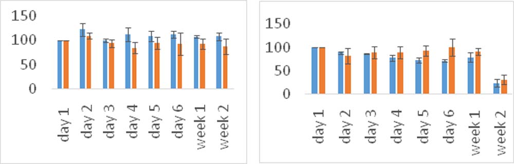
Metabolic Activity of Untreated and Treated Metabolic Activity of Untreated and Treated Normal Normal Epithelial Cells stromal cells TreatedUntreated
(b)Live dead assay:Intracellular esterase activity of live cells converts non-fluorescent cell-permeant calcein to intensely green fluorescent calcein. The Eth-D homodimer enters cells with damaged membranes, binds to nucleic acids, and produces a bright red fluorescence in dead cells.
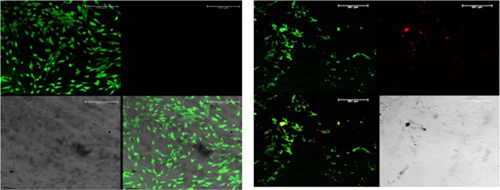
Untreated cells from limbal scleral explanttreated cells from limbal scleral explant
(2) Cross-linker penetration assay into human cornea :Fluorescently labelled N-hydroxysuccinimide (NHS-CF) was used to determine the extent of penetration in to the cornea. We used an ex-vivocorneal model to assess the penetration. Accordingly, donor corneas were washed with PBS and the endothelial side was plugged with agarose. The fluorescent cross-linker (either 20mM or 10mM) was applied to theintact epithelialside for 15 minutes in a CO2 incubator. Later, the corneas were washed and 8mm cryosections were taken and further imaged with a confocal microscope.
20mM NHS-FITC, 20x objective
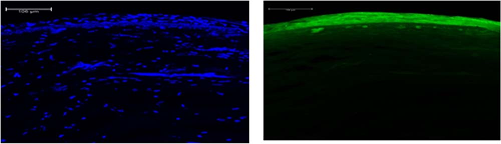
DAPINHS-FITC
(3)Tensile test analysis:After treatment with the crosslinker, the cornea are cut into strips with the help of customized mechanical punches. The size of the cut portion is 10mm length and 2mm breadth for the donor corneas and 7mm length and 2mm breadth for the keratoconic corneas. The instrument used to test the mechanical strength is the Universal testing machine Instron 3345 – Vecomtech using software instronbluehill lite, with 100μm/s strain rate, 50N load cell. The tensile strength was compared between the different groups of corneas.
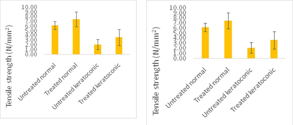
74% increase in tensile strength 4X increase in stiffness (young’s modulus)
CONCLUSIONS:
The novel cross-linker is able to induce the corneal stiffening of the weak keratoconic corneas while causing lesser cytotoxicity to the cells. Suberic acid is an emerging alternative to C3R for corneal crosslinking and has the potential to be further translated into a therapeutic agent. Further studies on endothelial cyototoxicity and dose modulation and will help us in understanding the bioavailability of suberic acid which will help us to proceed further with animal trials.
Leave a Comment