
Dr. Hrita Gogate, H19240, Dr. Hemanth Murthy, Dr. Muralidhar N S
Abstract:
The purpose of this study was to compare the outcomes of intraoperative 360° versus limited endolaser retinopexy in primaryMicro Incision Vitrectomy Surgery (MIVS) with gas for Rhegmatogenous Retinal Detachment (RRD). It was a retrospective, comparative, interventional study. Data of 89 eyes of 89 patients who underwent primary MIVS for RRD with intraoperative 360° laser (Group I) or limited laser (Group II) with 10 % C3F8 gas from January 2014 to December 2016 who fulfilled the inclusion and exclusion criteria were analysed. Main outcome measures were anatomical success rates and functional outcomes in both groups. Follow up period ranged from a minimum of 4 months to 9 months.62 patients underwent 360° laser (Group I) and 27 patients underwent localised laser retinopexy (Group II) out of which 60 were male and 29 were female with mean age of 56.75 years (range 16 to 87 years). Anatomical success rate was 90.32% in group Iand 96.3% in group II with no statistically significant intergroup difference (p>0.05). LogMAR BCVA improved from a mean of 1.769 to 0.679 in group I(p<0.05) and from 1.859 to 0.674 in group II (p<0.05) with no statistically significant intergroup difference (p>0.05). It was thus observed that 360° endolaser does not give any additional benefit over localised laser retinopexyin vitrectomy for RRD.
Introduction:
Rhegmatogenous retinal detachment (RRD) is a potentially blinding ocular condition. The prevalence of RRD ranges from6.3 to 17.9 per 100,000, with the highest incidence found in patients between 60 and 69 years of age1. The majorsurgical options for RRD repair include scleral buckling and pars plana vitrectomy. Recent advances in instrumentation, wide angle viewing systems and the use of various intra-operative tamponade agents have made vitrectomy procedures safer and preferred treatment. The surgery aims to identify and close all the retinal breaks with minimum iatrogenic damage. Vitrectomy consists of thorough vitreous removal, flattening of the retina and drainage of the subretinal fluid followed by creation of a strong retinochoroidaladhesion around the breaks. Thisis commonly achieved with the help of either cryotherapy or laser photocoagulation. However, cryotherapy may cause extensive breakdown of the blood retinal barrier, enhance the release of viable RPE cells and thus result in an increase in subsequent proliferative vitreoretinopathy (PVR). Laser photocoagulation is used widely since it rapidly induces retinochoroidal adhesion and has lesser tendency to cause PVR. Laser can be applied in a 360° manner or just limited to the retinal breaks and other suspicious areas. The quantum of laser energy may also have an effect in the tendency to cause PVR. The main purpose of this study was to analyse the anatomical and functional outcomes of intraoperative 360° versus limited endolaser retinopexy in primary micro incision vitrectomy surgery (MIVS) with gas for rhegmatogenous retinal detachment (RRD).
Methods:
Patients:
The medical records of patients operated for RRDs from January 2014 to December 2016 at Retina Institute of Karnataka were reviewed. All patients who had RRDand underwent primary MIVS with either intraoperative 360° or localised endolaser retinopexy were included. The exclusion criteria were (1) a prior history of vitreoretinal surgery; (2) PVR worse than grade C1; (3) other severe vision-impaired eyediseases (e.g., advanced glaucoma or macular hole); (4) non-primary RRD such as recurrent RD, and RD associated withuveitis or diabetes mellitus, and (5) giant retinal tears (6) minimum follow-up period<4 months. The review yielded 89 eyes of 89 patients,out of which 62 had undergone 360° endolaser( group I) and 27 had undergone localised endolaser retinopexy (group II).
Data:
Age, gender, extent of RD, characteristics of the break such as number of breaks(single or multiple) and type of breaks(hole or horseshoe tear), macular and lens status, pre- and postoperative best-corrected visual acuity (BCVA), intraocular pressure (IOP) and the complications encountered were noted. The eyes were analysed in two groups: 360° laser retinopexy (group I) and localised laser retinopexy (group II). The decision about the extent of laser application was made intraoperatively based on the surgeon’s preference and the intraoperative retinal condition. Written informed consent was obtained from all patients before surgery.
Surgical details:
All the procedures were performed under peribulbar anesthesia. Three-portMIVS was performed using the Alcon Constellation system (AlconLaboratories, Inc., Fort Worth, TX, USA). First,core vitrectomy was performed. Then, in eyes without a pre-existing posterior vitreous detachment (PVD), triamcinolone acetonide assisted PVD induction was done. Following this, a thorough peripheral vitreous base trimming was done with scleral depression.Retina was flattened using either air or perfluorocarbon liquid (PFCL) till the posterior edge of the break.360° scleral depression was performedagain to check for residual retinal breaks and areas of degeneration.Complete fluid air exchange or PFCL-air exchange (when PFCL was injected) was performed. Three rows of confluent medium-white laser burns wereperformed with a 532-nm laser (power range 200 mW, duration 100ms) using a direct laser delivery system (Purepoint laser). In group I, laser photocoagulation was applied within two optic disc diameters posterior to the ora serrata. In group II, laser was applied around the break and to other suspicious areas. Subsequently, nonexpansile gas(10% Octafluoropropane (C3F8)) was used for endotamponade. All the patients were asked to maintain a specific head posture (depending on the location of break) for 7-14 days. Patients were discharged on the same day and were called for review on postoperative day 1, week 1,week2, 1 month, and then bimonthly till a minimum of 4 months in the postoperative period. Anatomically successful surgery was defined as the complete disappearance of SRF and flattening of the retina. Functional outcome was determined by comparing the pre operative and post operative BCVA. Complications, if any, were noted.
Statistical analysis:
BCVA was converted to the logarithm of the minimum angle of resolution (logMAR). Hand motion, and light perception were assigned as a logMAR unit of 2, and 3 respectively. First, demographic and preoperative clinical indicators of two groups were noted. Post operative parameters were analysed using either Student’s t test (continuous factors) or a chi-square test (categorical factors). P value <0.05 was considered statistically significant.
Results:
A total of 89 eyes of 89 patients were included in this study. 62 eyes underwent 360° endolaser and were included in group I and 27 eyes underwent localised endolaser retinopexy and were included in group II. The follow-up period ranged from a minimum of 4 months to 9 months with a mean of 6.21 months. Demographic and baseline clinical characteristics of the two groups of patients are summarized in Table 1. All the eyes achieved statistically significant improvement in BCVA after surgery (p<0.05). Anatomical success rate (Table 2) was 90.32% in group I and 96.3% in group II with no statistically significant intergroup difference (p>0.05). LogMAR BCVA improved from a mean of 1.769 to 0.679 in group I (p<0.05) and from 1.859 to 0.674 in group II (p<0.05) with no statistically significant intergroup difference (p>0.05){Table 3 and Figure 1}. 6 eyes in group I and 1 eye in group II had redetachment which was treated with revision vitrectomy and silicon oil endotamponade. 2 eyes developed cataract in each group during the course of follow up for which they underwent phacoemulsification with IOL. 15 eyes in group I and 7 eyes in group II had rise in IOP which was managed with topical antiglaucoma medications. 1 eye in group I developed subretinal fibrosis in the inferior quadrant but it was only observed since it did not warrant any treatment. Complications encountered are enlisted in Table 4.
Table 1: Demographic and other pre-operative characteristics
| Parameter | Total | Group I | Group II |
| Mean Age
(years) |
56.75
(ranged from 16 to 87) |
55.85
(ranged from 16 to 85) |
59.44
(ranged from 32 to 87) |
| Gender
Male Female |
60 29 |
41 21 |
19 8 |
| Extent of RD
≤ 6 clock hours > 6 clock hours |
46 43 |
27 35 |
19 8 |
| Number of breaks
Single Multiple |
64 25 |
44 18 |
20 7 |
| Type of break
Only retinal hole Only horseshoe tear Both |
14 70 5 |
9 48 5 |
5 22 0 |
| Macular status
On Off |
20 69 |
17 45 |
3 24 |
| Lens
Phakic Pseudophakic Aphakic |
31 56 2 |
20 40 2 |
11 16 0 |
Table 2: Anatomic outcomes
| Group I | Group II | |
| Anatomical success | 56 (90.32%) | 26 (96.3%) |
| Redetachment | 6 (9.68%) | 1 (3.7%) |
Table 3: Comparative pre operative and post operative best corrected visual acuity(BCVA) in both groups
| Group I | Group II | P value | |||
| Mean pre operative BCVA |
1.769 |
1.859 |
0.7233 | ||
| Mean post operative BCVA | 0.679 | 0.674 | 0.9632 | ||
| Mean change in BCVA | 1.063 | 1.185 | 0.5928 |
Figure 1: Comparative pre operative and post operative best corrected visual acuity (BCVA)
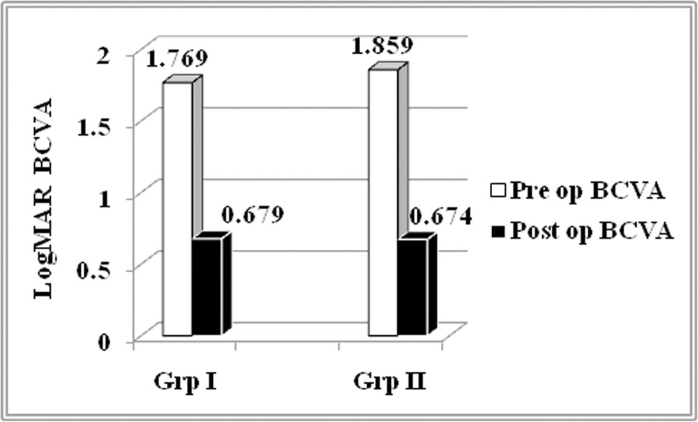
Table 4: Complications
| Complication | Group I | Group II |
| Rise in IOP | 15 | 7 |
| Cataract | 2 | 2 |
| Subretinal fibrosis | 1 | 0 |
Discussion:
Scleral buckling (SB) and pars plana vitrectomy (PPV) are the two main surgical treatments for RRD. Vitrectomy was found to be more efficacious earlier in pseudophakic eyes 2-4, and many surgeons preferred SB in younger phakic RRD without posterior vitreous detachment (PVD) and those with less-liquefied, formed vitreous 5,6. However, clinical trials have showed comparable efficacy between the two surgical procedures for treating uncomplicated phakic RRD 2,5,7,8. Choice of procedure is now determined by the surgeon’s discretion in uncomplicated phakic RRD. The safety and effectiveness of PPV has improvedowing to small gauge systems, better instrumentation and wide angle viewing system (WAVS) 9. The WAVS enhances surgical procedures, especially for conditions like RD, because it provides detailed information about peripheral retina.
The main aim of vitreoretinal procedures in management of RRD is to accurately localise all retinal breaks and to seal them (retinopexy). This is achieved by cryotherapy or by endolaser. However, cryotherapy achieves retinochoroidal adhesion later than laser photocoagulation. Also, cryotherapy causes more severe breakdown of the blood retinal barrier and more release of RPE cells and other inflammatory mediators,thus increasing the risk of subsequent proliferative vitreoretinopathy (PVR)
Laser photocoagulation is used to treat localised RD and retinal breaks because it can rapidly enhance retinochoroidal adhesion in vitro and in vivo to 140% of normal within 24 h and three times that of normal at 2 weeks 10. For laser photocoagulation to be effective, an adequate extent and optimum placement of the laser are of great importance. Although lesser than cryotherapy, excessive laser photocoagulation has been implicated as a risk factor for future PVR. Hence, judicious use of laser is vital.
This study aimed at comparing anatomic and functional outcomes of localised retinopexy versus 360° retinopexy to determine whether additional laser augments retinochoroidal adhesion or affects the incidence of PVR. As evident from the results, we found that success rates were 90.32% in group I and 96.3% in group II. Also, both groups showed statistically significant improvement in BCVA. However, the intergroup difference was statistically insignificant (p>0.05). Thus, 360° laser did not provide any additional benefit over localised laser retinopexy.
Quantum of laser photocoagulation has been studied previously in cases of RRDs with undetected breaks. Zhou et. al.11 compared the efficacy of 360° and localised laser retinopexy in PPV for the management of RRD with intraoperative undetected breaks. The results of this study indicated that the eyes that underwent 360° laser retinopexy achieved a higher primary success rate and greater improvement in postoperative BCVA in cases of undetected breaks. These results were coherent with prior studies which have consistently noted that the tears detected in RRDs with unseen retinal breaks were mostly small anterior tears located in the far periphery of retina 12-14. Therefore, localised laser photocoagulation probably could miss these innocuousbreaks.
However, there are no definite guidelines in literature to accurately quantify the amount of laser applications required or to justify 360° laser retinopexy as compared to localised retinopexy in RRDs with well localised breaks. This study, therefore, should be regarded as an initial attempt at analysing results of the different applications of laser applied in MIVS for RRD with well localised breaks.
However, caution should be exercised when interpreting the findings due to several limitations. Firstly, the main drawback of this study was its retrospective and nonrandomized nature. Secondly, the sample size was small, and all the patients were recruited from a single tertiary institution. Also, the decision about the quantum of laser application was determined by the surgeon’s preference. These factors might have caused selection bias, and our results cannot be necessarily extrapolated to the entire RRD population.It is, however, likely that limited laser retinopexy would yield benefits in reduced operating time, lower post operative morbidity and complications like raised IOP and PVR.
Overall, compared with localised laser retinopexy, 360° laser retinopexy did not give any additional benefit with respect to anatomic and visual outcomes for RRD patients. It does, however, require a randomized, prospective trial with systematic long term follow-up to fully validate the findings of this study.
Financial disclosure:
No author has any financial interest in any procedure or product used.
References:
- D.Mitry, D.G. Charteris, B.W. Fleck,H. Campbell, and J. Singh, “The epidemiology of rhegmatogenous retinal detachment: geographical variation and clinical associations,” British Journal of Ophthalmology, vol. 94, no. 6, pp. 678–684, 2010.
- Heimann H, Bartz-Schmidt KU, Bornfeld N, Weiss C, Hilgers RD, Foerster MH, et al. Scleral buckling versus primary vitrectomy in rhegmatogenous retinaldetachment: a prospective randomized multicenter clinical study.Ophthalmology. 2007;114(12):2142–54. doi:10.1016/j.ophtha.2007.09.013.
- Sharma YR, Karunanithi S, Azad RV, Vohra R, Pal N, Singh DV, et al.Functional and anatomic outcome of scleral buckling versus primaryvitrectomy in pseudophakic retinal detachment. Acta Ophthalmol Scand.2005;83(3):293–7. doi:10.1111/j.1600-0420.2005.00461.
- Brazitikos PD, Androudi S, Christen WG, Stangos NT. Primary pars planavitrectomy versus scleral buckle surgery for the treatment of pseudophakicretinal detachment: a randomized clinical trial. Retina. 2005;25(8):957–64.
- Miki D, Hida T, Hotta K, Shinoda K, Hirakata A. Comparison of scleralbuckling and vitrectomy for retinal detachment resulting from flap tears insuperior quadrants. Jpn J Ophthalmol. 2001;45(2):187–91.
- Schneider EW, Geraets RL, Johnson MW. Pars plana vitrectomy withoutadjuvant procedures for repair of primary rhegmatogenous retinaldetachment. Retina. 2012;32(2):213–9. doi:10.1097/IAE.0b013e3182278b29.
- Koriyama M, Nishimura T, Matsubara T, Taomoto M, Takahashi K, MatsumuraM. Prospective study comparing the effectiveness of scleral buckling tovitreous surgery for rhegmatogenous retinal detachment. Jpn J Ophthalmol.2007;51(5):360–7. doi:10.1007/s10384-007-0463-0.
- Azad RV, Chanana B, Sharma YR, Vohra R. Primary vitrectomy versusconventional retinal detachment surgery in phakic rhegmatogenous retinaldetachment. Acta Ophthalmol Scand. 2007;85(5):540–5. doi:10.1111/j.1600-0420.2007.00888.
- Chalam KV, Shah VA. Optics of wide-angle panoramic viewing systemassistedvitreous surgery. Surv Ophthalmol. 2004;49(4):437–45. doi:10.1016/j.survophthal.2004.04.010.
- Yoon YH, Marmor MF (1988) Rapid enhancement of retinal adhesionby laser photocoagulation. Ophthalmology 95(10):1385–1388.
- Chuandi Zhou & Zhi Zheng & Qinghua Qiu. Pars plana vitrectomy with 360° versus localised laser retinopexy in the management of retinal detachment with undetected breaks intraoperatively: a retrospective, comparative, interventional study . Lasers Med Sci (2017) 32:583–589.DOI 10.1007/s10103-017-2152-7.
- Brazitikos PD, D’Amico DJ, Tsinopoulos IT, Stangos NT (1999) Primary vitrectomy with perfluoro-n-octane use in the treatment of pseudophakic retinal detachment with undetected retinal breaks. Retina 19(2):103–109.
- Kita M, Yoshimura N (2011) Endoscope-assisted vitrectomy in the management of pseudophakic and aphakic retinal detachments with undetected retinal breaks. Retina 31(7):1347–1351.
- Martinez-Castillo V, Boixadera A, Garcia-Arumi J (2009) Pars plana vitrectomy alone with diffuse illumination and vitreous dissection to manage primary retinal detachment with unseen breaks. Arch Ophthalmol 127(10):1297–1304.
Leave a Comment