
Dr. Jay Vardhan, V19035, Dr. Mohan Shalini, Dr. Sachan S.K., Dr. Mohit Khattri
INTRODUCTION:
Penetrating keratoplasty is a widely used procedure even today in spite of various recent advances in corneal surgeries.
Corneal buttons are achieved after host cornea trephination following penetrating keratoplasty. Histopathological study of corneal button is extremely valuable to analyses the degree and extent of corneal involvement which may show specific histological pattern and may aid in management of various condition.1-5
The study was done with the aim of correlating clinical diagnosis with histopathological diagnosis.6
MATERIAL AND METHOD:
This was a retrospective study done at a tertiary care hospital. The records of patients who underwent penetrating keratoplasty from 01April 2015 to 31 March 2016 were reviewed. The patients whose histopathological reports were available, were included in the study. This lead to inclusion of 93 patients in the study. All corneas were handled in accordance with the guidelines of the Declaration of Helsinki regarding research on human tissue. Informed consent was checked from all subjects that there was no risk involved in donating their tissue. Detailed history and examination was reviewed with emphasis on the details of the corneal button sent for histopathology was studied in detailed. In case of any doubt the histopathological section was again reviewed with the help of ahistopathologist. The findings of both light microscopy and electron microscopy was studied in details. The tissue section was examined at the magnification power of 100x & 400x.
RESULTS:
Out of 93 patients, 61 were in age group 51-70 year age group;36 of them were male and 25 were female (Table 1). Histopathologically, 52 out of 56 patients of bullous keratopathy,12 out of 14 patient of corneal opacity;4 out of 8 patients of graft rejection and all patients of corneal ulcer/keratitis with or without perforation / descematocele and corneal degenerations were in correlation with clinical diagnosis.
Various histopathological changes seen in the corneal button were analyzed:
Corneal opacity:On histology of these opacity various changes seen are tabulated in Table 2 (Figure 1). Two out of 14 patients had bullae formation seen along with corneal opacity. Bowman’s membrane was thickened, distorted and with or without splitting .In scar condition Bowman’s membrane may or may not be intact . Stroma showed increased thickness in majority alongwith sclerosis in 2 patients.
Corneal degenerations: Corneal degeneration was noted in patients.
One patients showed thinned out epithelium and at places separation of epithelium from Bowman’s layer created spaces. There were spheroidal degeneration changes within the epithelium (Figure 2). Bowman’s layer and superficial stroma also showed spheroidal changes.
Second patient showed attenuated epithelium. Subepithelial tissue comprising of fibrocellular tissue which replaced the Bowman’s membrane .Underlying stroma showed scarring and the basement layer was distorted .Few unevenly distributed keratinocytes were present. The findings were suggestive of Salzmann’s nodular degeneration.
Corneal ulcer and Desmatocele:- Squamousepithelium was disrupted in continuity with underlying stroma showed loosened lamellae (Table 3). Mild inflammation with inflammatory cells alongwith stromal fibrous proliferation was also seen.(figure-3,figure-4)
Graft rejection: Eight patients showed infilteration of lymphocytes and plasma cells alongwith proliferation (Figure 5, Table 4).
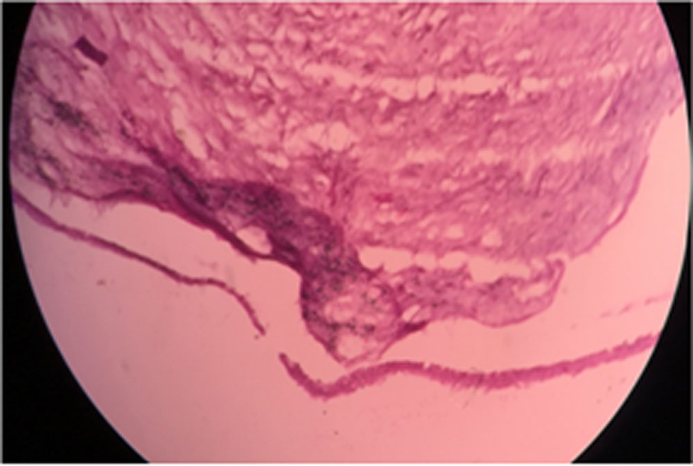
Bullous keratopathy :- The squamous epithelium showed separation from bowman’s layer creating bullous detachment . There were hydropic change within the epithelium (table 5) .
DISCUSSION :
On histological examination of established clinical diagnosis maximum number of corneal specimen resembles to their diagnosis.
A Meeney , HS Mudhar et al6showed in their study that in 44/50 (88%) of cases, histologicaldiagnosis matched the consultant’s diagnosis in first 6 months.In next 24 months, 48/50 (96%) of cases, histologicaldiagnosis matched the consultants’ diagnosis.
Histology of corneal opacity shows similarity with PK Agarawal et al (1983)12 , corneal ulcer & bullous keratopathy Geeta K et al 11.
CONCLUSION:1.On histopathological studyof host corneal button contributes to clinical management especially in case of corneal infections.
- In graft failure histology shows extent of host cornea involvement and further prognosis.
- Evaluation of clinical diagnosis can be done by histology.
Conflict of interest: No conflict of interest.
REFERENCES:
1:Jakits, M. A.: The fine structure of the human cornea, in Smelser, G. K., editor: The structure of the eye, New York, 1961, Academic Press, Inc., p. 343.
2.Huang AJ, Wichiensin P, Yang M. Bacterial keratitis. In: Krachmer JH, Mannis MJ, Holland EJ,editors. Cornea: Fundamentals, Diagnosis and Management. Vol. 1. Elsevier-Mosby; 2005. pp. 1025–33. Ch. 81.
3.Vemuganti GK, Garg P, Gopinathan U, Naduvilath TJ, John RK, Buddi R, et al. Evaluation of agent and host factors in progression of mycotic keratitis: A histopathological and microbiological study of167 corneal buttons. Ophthalmology. 2002;109:1538–46.
4.Eagle RC. Eye Pathology an Atlas and Basic Text. WB SaundersCompany: Philadelphia, 1999.
5.Lee WR. Ophthalmic Histopathology. 2nd edn. Springer-Verlag:London, 2002
6.A Meeney and HS Mudhar: Histopathological reporting of corneal pathology by a biomedical scientist: the Sheffield Experience:2013
7.Geeta K. Vemuganti, Somasheila I. Murthy, and Sujata Das. Update on Pathologic Diagnosis of Corneal Infections andInflammations : 2011
8.PK Agrawal :The pathology of cornea (A histopathological study) :1983
Legends:
Figure-1: Histopathological features of Leucomatous corneal opacity
Figure-2: Histopathological features of Spheroidal degeneration
Figure 3: Histopathological features of Superficial corneal ulcer
Figure-4: Histopathological features of Desmatocele
Figure-5: Histopathological features of Bullous keratopathy
Table -1: Demographic distribution of study participants
Table-2 : Histopathological findings in Corneal opacity & degeneration
Table-3: Histopathological findings in Corneal ulcer & keratitis
Table-4: Histopathological findings in Graft rejection
Table-5: Histopathological findings in Bullous keratopathy
FIGURES :
Figure-1: Histopathological features of Leucomatous corneal opacity
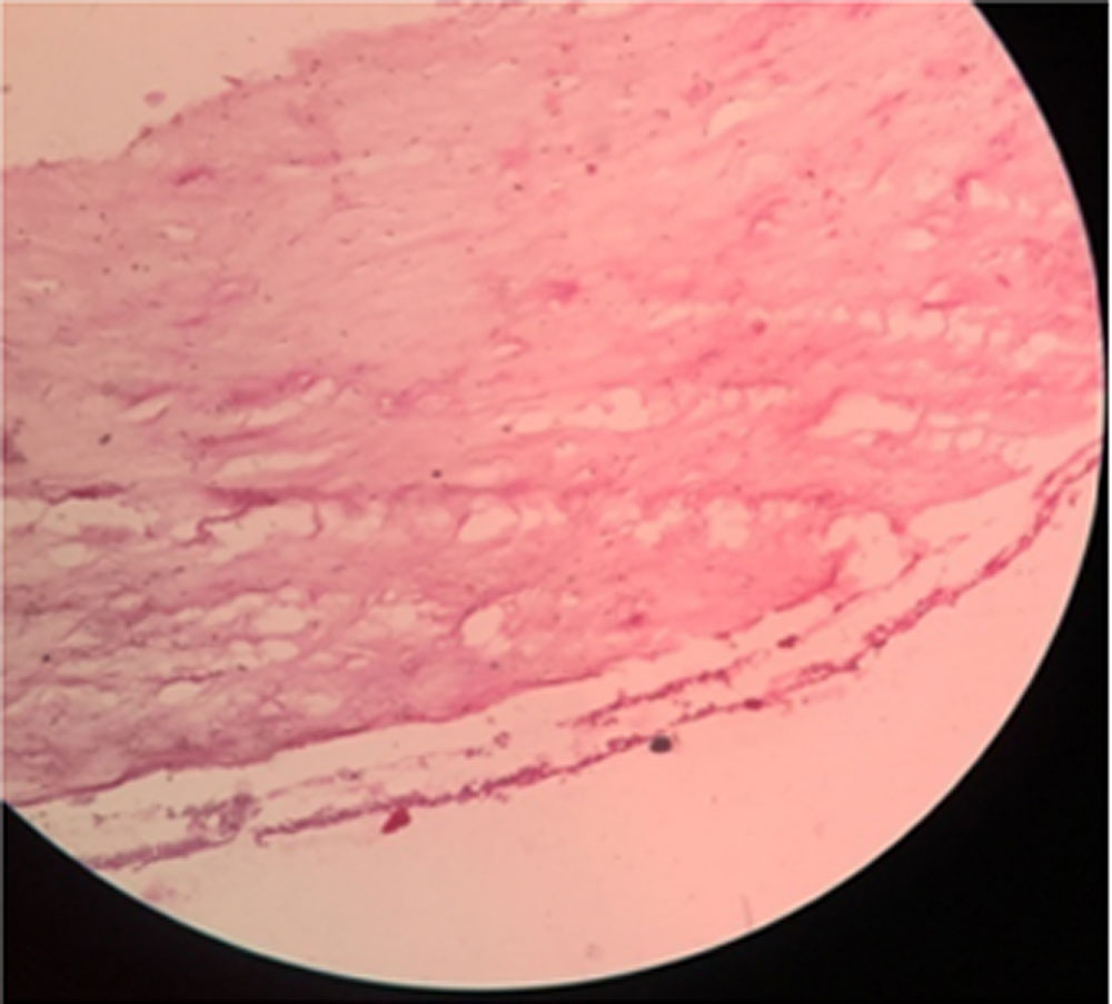
Figure-2: Histopathological features of Spheroidal degeneration
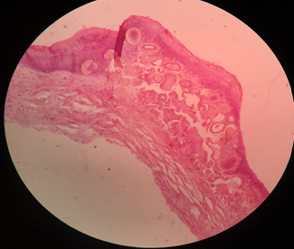
Figure 3: Histopathological features of Superficial corneal ulcer
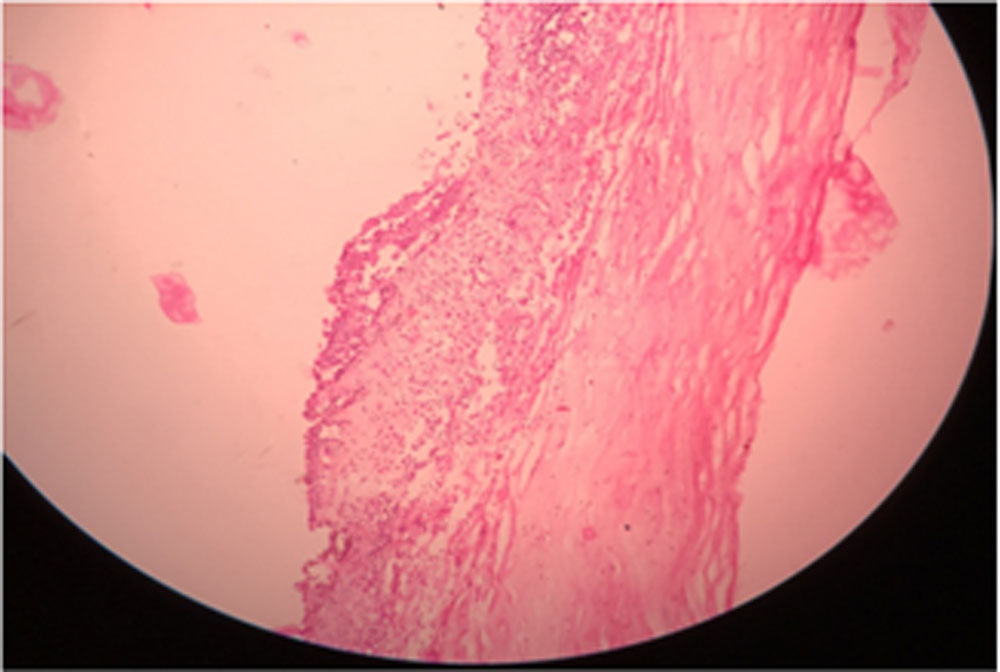
Figure-4: Histopathological features of Desmatocele
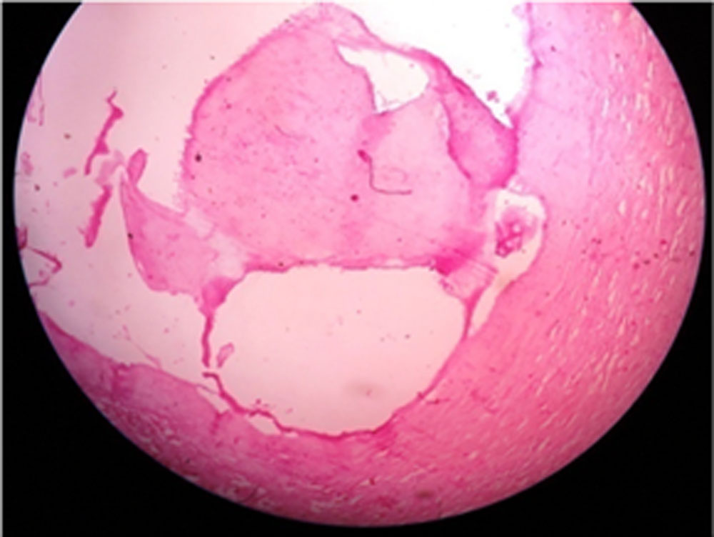
Figure-5: Histopathological features of Bullous keratopathy
Tables :
Table -1: Demographic distribution of study participants
| Age group | Male | Female | Total |
| 21-30 | 0 | 2 | 2 |
| 31-40 | 6 | 4 | 10 |
| 41-50 | 5 | 6 | 11 |
| 51-60 | 20 | 13 | 33 |
| 61-70 | 16 | 12 | 28 |
| 71-80 | 7 | 2 | 9 |
| 54 | 39 | 93 |
Table-2 : Histopathological findings in Corneal opacity& degeneration
|
Layer of cornea |
Histological changes in No. of specimen out of 14 corneal opacity& 2 Degenerations |
|||||
|
Epithelium |
Continuous | Atrophic | Hyperplasia | Separation from BM | Bullae formation | Degeneration |
| 10 | 1 | 1 | 2 | 2 | 2 | |
|
Bowman’s membrane (BM) |
Normal | Thickening | No comments | |||
|
0 |
Splitting | Without splitting | 11 | |||
| 2 | 1 | |||||
|
Stroma |
Increased thickness | Irregular
thickness |
Sclerosis | Vascularisation | Degeneration | |
| 12 | 4 | 2 | 4 | 2 | ||
|
Descemet’s membrane |
Intact | Perforation | Coiling | Duplication | Disruption | |
| 7 | 2 | 3 | 2 | 2 | ||
|
Endothelium |
Intact | Loss | Atrophic | Degeneration | ||
| 8 | 1 | 4 | 1 | |||
Table-3: Histopathological findings in Corneal ulcer & keratitis
| Layer of cornea | Histological changes | No. of specimen out of 13 corneal opacity button | |
| Epithelium | Breach in continuity | 13 | 0 |
| Bowman’s membrane | Disruption | 13 | 0 |
|
Stroma |
Inflamed | 13 | 0 |
| Stromal edema | 10 | 3 | |
| Infiltration of inflammatory cell | 13 | 0 | |
| Stromal proliferation | 8 | 5 | |
| Descemet’s membrane | Intact | 11 | 2 |
| Protrusion | 3 | 10 | |
| Incarcetion in stroma | 6 | 7 | |
| Endothelium | Intact | 10 | 3 |
Table-4: Histopathological findings in Graft rejection
| Layers of cornea | Histological changes in
No. of specimen out of 8 corneal opacity button |
||
| Epithelium
|
Intact | Breeched
|
Cellular infiltrates |
| 3 | 5 | 4 | |
| Stroma | Edema | Cellular infiltrates
|
|
| 6 | 4 | ||
| Endothelium | Swelling | Cellular infiltrates | Loss/ discontinuation |
| 3 | 3 | 3 | |
Table-5: Histopathological findings in Bullous keratopathy
| Layer of cornea | Histological changes
No. of specimen out of 56 corneal button |
|||
| Epithelium | Intact | Bullae formation | Hydropic changes | |
| 52 | 54 | 50 | ||
| Bowman’s membrane | Thinning | 4 | ||
| Stroma | Irregular Lamellae pattern | Stromal edema | ||
| 49
|
56 | |||
| Descemet’s membrane | Intact | 56 | ||
| Endothelium
|
Attenuated | Discontinued | ||
| 52 | 4
|
|||
Leave a Comment